STUDY OF PREVALENCE ON CORAL BLEACHING AND DISEASES
on
ECOTROPHIC • 5 (2) : 123 - 128
ISSN: 1907-5626
STUDY OF PREVALENCE ON CORAL BLEACHING AND DISEASES
EGHBERT ELvAN AMPou*
*Ministry of Marine Affairs and Fisheries - Institute for Marine Research and Observation Marine Conservation Research 'Team, Bali, Indonesia
E-mail: elvan_ampou76@yahoo.com; elvan_ampou@dkp.go.id
ABSTRAK
Monitoring nilai prevalence karang yang mengalarni pemutihan clan penyakit sangat perlu dilakukan secara intensiv di Indonesia yang juga masuk dalam kawasan CTI, informasi tentang hal ini boleh dikatakan relatif belum banyak dilakukan orang. Metode yang dipakai selama survey adalah time swim dimana dibagi pada 2 kedalaman (S clan 10 meter) selama 30 menit, untuk analisis data digunakan rumus prevalence. Nilai Prevalence karang yang mengalarni pemutihan clan penyakit di Raja Ampat pada kedalaman Sm=30,67%; 10m=23,50% di bulan November 2009. Taman Nasional Bunaken kedalaman Sm=SS,47%; 10m=83,73% di bulan Agustus 2009 clan di Pulau Runduma-Taman Nasional kedalaman Sm=23,55%; lOm=S0,94% di bulan Oktober 2009.Jenis karang yang dominan mengalami pemutihan clan penyakit adalah genus Porites clan Acropora, sedangkan tidak dominan adalah genus Pocillopora clan Montipora.
Kata kunci: pemutihan karang, time swim, prevalence.
INTRODUCTION
Coastal zone and Indonesian seas have a potential and high biodiversity (mega biodiversity) in the world also include in CTC (Coral Triangle Center) region. The highly biodiversity comprise in genetic, species and other ecosystem to develop Indonesia economics, environment, sustainability and carrying capacity also Law reinforcement (Anonimous, 2007).
Based on characterize coastal ecosystem is natural or man made. The natural ecosystem located on the coastal zone, ex: coral reefs, mangrove forest, seagrass beds, sandy beach, barringtonia formation, estuary, lagoon, delta and small island ecosystem. (Dahuri, 2003).
Coral diseases and syndromes generally occur in response to biotic stresses such as bacteria, fungi and viruses, and/or abiotic stresses such as increased sea water temperatures, ultraviolet radiation, sedimentation and pollutants. One type of stress may exacerbate the other (Santavy and Peters, 1997).
Major Reef-Building Coral Diseases
The frequency of coral diseases appears to have increased significantly over the last 10 years, causing widespread mortality among reef-building corals. Many scientists believe the increase is related to deteriorating water quality associated with anthropogenic pollutants and increased sea surface temperatures. This may, in tum, allow for the proliferation and colonization of disease-causing microbes. However, exact causes for most coral diseases remain elusive. The onset of most diseases likely is a response to multiple factors (Peters, 1997).
This section discusses, in alphabetical order, the
most prevalent coral diseases and syndromes currently known and under study: black-band disease, coral bleaching, dark-spots disease, red-band disease, whiteband disease, white-plague disease, white pox and yellow-blotch disease. Additional information on these diseases and others can be found on NOAA's Coral Disease Identification and Information Web site.
Coral Bleaching
Healthy tissue of most stony corals ranges from yellow to brownish in color, a function of the photosynthetic pigments of their symbiotic zooxanthellae. When corals are inordinately stressed, they often expel their zooxanthellae, or the concentration of photosynthetic pigments declines. This response is known as bleaching (Glynn, 1996).
During a bleaching event, a coral's coloration disappears or becomes pale, and the white of the coral skeleton shows through the translucent coral tissue.In some species, such as the massive starlet coral Sideras-trea sidereal, the tissue can appear pinkish or bluish, due to pigments within the animal tissue. Localized bleaching has been observed since at least the beginning of the 20th century. However, beginning in the 1980s, regional and global bleaching affecting numerous species has occurred on reefs worldwide. Bleaching usually is not uniform over single coral colonies within coral communities or across reef zones, and some species are more susceptible to bleaching than others under the same conditions (Glynn, 1996). In some instances, only the upper surface or lower surface of the colony is affected. In others, bleached tissue appears as a circular patch or in the shape of a ring or wedge.
Localized bleachinghas been attributed to exposure to high light levels, increased ultraviolet radiation, tern-
perature or salinity extremes, high turbidity and sedimentation resulting in reduced light levels, and other abiotic factors (Glynn, 1996). In addition, bleaching in some species has occurred in response to a bacterial infection (Kushmaro et al., 1996). However, the seven major episodes of bleaching that have occurred since 1979 have been primarily attributed to increased sea water temperatures associated with global climate change and el Niii.o/la Nina events, with a possible synergistic effect of elevated ultraviolet and visible light (Hoegh-Guldberg, 1999).
Debilitating effects of bleaching include reduced skeletal growth and reproductive activity, and a lowered capacity to shed sediments and resist invasion of competing species and diseases (Glynn, 1996). Prolonged bleaching can cause partial to total colony death. If the bleaching is not too severe, and the stressful conditions decrease after a short time, affected colonies can regain their symbiotic algae within several weeks to months (Glynn, 1996).
One predicted effect of climate change is increased coral bleaching (whitening), which is caused by the disruption of the symbiotic relationship between polyps and zooxanthellae resulting in the expulsion of zooxanthellae and loss of photosynthetic pigments. Stresses that can cause this include freshwater flooding, pollution, sedimentation, disease and, most importantly, changes in light and temperature. If stresses continue for long enough, corals and whole reefs can suffer reduced fecundity and growth rates, and eventually even mortality. Once sections of the coral reef die they become vulnerable to further structural degradation by algal overgrowth and bioerosion. Overall, though, the bleaching phenomenon is extremely patchy and can vary greatly according to location, environmental conditions, season or species composition. (Douglas, 2003).
Based on the background, the aims of this research are 1) To Investigate about prevalence on coral bleaching and diseased, and 2) To determine dominant of the genus/species experience of bleaching and diseased.
METHODS
Study Area
Research location was consisted in 3 sites: TN Bu-naken (North Sulawesi) inAugust 2009, TN. Wakatobi (Southeast Sulawesi) in October 2009 and KKP Raja Ampat (Birdshead of Papua) in November 2009.
Time Swims
Diver swim with given time. In this survey used 30 minutes to collect data especially bleaching/diseases and dive in 2 depths (5 and 10 meters).
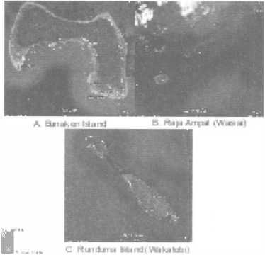
Figure 1. Survey location map from 3 site study to support data validation. A. Bunaken Island as the one part of Bunaken National Park-North Sulawesi., B. Marine Protected Area are so called Kawasan Konservasi Perairan (KKP) in Raja Ampat (Waisai) Bird's Head of Papua C. Runduma Island northeast from Wakatobi district as the one region of Wakatobi National Park-Southeast Sulawesi. (Google map 2010).
Diseased Prevalence
Diseased Prevalence is the proportion of diseased colonies to the total measured population of colonies. It can be calculated for individual populations, species or genera, or for the coral community as a whole, as well as for each particular disease/syndrome, similar group of diseases or for all diseases lumped together. What is calculated depends on the question asked.
-
- Prevalence(P) = (# diseased colonies/total # colonies) x 100
-
- Total Prevalence (P) = (# diseased colonies/total # colonies) x 100 / I Location
A prevalence value is estimated for each area-sample unit. An average prevalence value with standard deviation can then be calculated for habitats, zones or reefs (depending on the stratification and the question) using the sample unit prevalence value (Coral Diseases handbook, 2008).
Table 1. Prevalence category coral bleaching and diseases
|
No |
Prevalence (%)∕location∕Genus-Spesies |
Category |
|
1 |
0-50 |
good |
|
2 |
51 -75 |
medium |
|
3 |
76-100 |
poor |
|
4 |
>100 |
PoorlyI serious condition |
RESULTS AND DISCUSSION
Bunaken Island
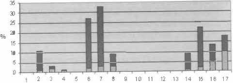
Total Prevalence at depth Sm 55,47%/Location/ Genus-Sp. The dominant bleaching & diseased is Po-rites sp and Acropora in Ron's point. Prevalence = 113,33% and Muka Kampung = 80%. Less Prevalence in Celah-celah = 7% and Lekuan 2 = 6,6%.
Total Prevalence on lOm = 83,73%/Lokasi/Ge-nus-Sp. Dominant bleaching & diseased average from genus :Acropora, Pocillopora and Montipora. Which are dominant it was occurred in Lekuan 2 especially species Symphyllia radians. With Prevalence = 133,33% . Whereas less 50% in Celah-celah=39,44%, Lekuan 1=33,71% dan Fukui=l6,66%.
Pre•Jalence 8 ea cli rg and Cl se ase d in 5m deJ:i h
Table. 2. Species Encountered in Sm depth
|
No |
Location |
Coordinates |
Genus/Seecies |
|
I |
Cclah-Celah |
N !' 35" 56.9 E 124' 46" 00.0 |
Acropora brueggema11i, Physogyra |
|
2 |
Lekuan 2 |
N !' 36" 58.0 E 124' 45" 54.0 |
Acropora palifera |
|
3 |
Muka Kampung |
N !' 35" 38.0 E 124' 46" 28.4 |
Porites sp, Pocillopora sp, Gonio-pora sp |
|
4 |
Lekuan l |
NJ' 35" 41.6 E 124' 46" 16.3 |
Porites mayeri |
|
5 |
Ron's Point |
N !' 36" 22.3 E 124' 44" 09.4 |
Porites rnayeri, Acropora brueggemanni,Goniastrea minuta, Goniastrea retiformis |
|
6 |
Fukui |
NI' 36" 44.l E 124' 44" 22.9 |
Oxypora lacera, Acropora prostate, Oxll'ora lacera |
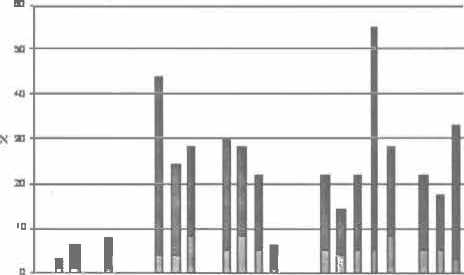
Figure 2. Prevalence in Sm
Table. 3. Species Encountered in Sm depth
|
No |
Location |
Coordinates |
Genus/Seecies |
|
l |
Celah-Celah |
N J' 35" 56.9 E 124' 46" 00.0 |
Mo11tipora foliosa, Montipora informis, Acropora nobilis, Acropora palifera |
|
2 |
Lekuan 2 |
NJ' 36" 58.0 E 124' 45" 54.0 |
Acropora palifera, Tubastrea mi-crantha, Symphyllia radians |
|
3 |
Muka Kampung |
NJ' 35" 38.0 E 124' 46" 28.4 |
Acropora palifera, Go,iiastrea reti-formis, Pocillopra verrucosa, Acrop-ora Formosa, Acropora millepora, Goniastrea minuta, Pocillopora verrucosa, Porites stephensoni |
|
4 |
LekuanI |
N l' 35" 41.6 E 124' 46" 16.3 |
Acropora millepora |
|
5 |
Ron's Point |
N l' 36" 22.3 E 124' 44" 09.4 |
Acropora palifera, Porites stephenso-ni, Stylophora pistillata, Pocillopor,1, Isopora sp |
|
6 |
Fukui |
N l' 36" 44.l E 124' 44" 22.9 |
Acropora yongei, Pocillopora verucosa, Montieora sf'! Porites lutea |
Prevalence B leaching and D iseased in 1 0 m
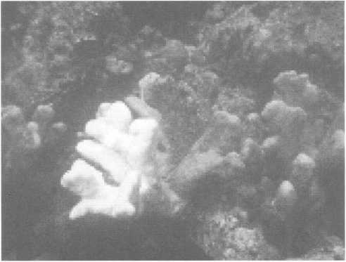
Figure 3. Acropora palifera at Celah-celah dive site at depth lOm with "unusual bleaching patterns"
RajaAmpat
Table. 4. Species Encountered in Sm depth
|
No |
Location |
Coordinates |
Genus/Seecies |
|
1 |
Saonek Monde (SN) |
unrecord |
Porites, Acropora (3x), Porites |
|
2 |
TanjungSaleo (TS) |
00° 26.475' 130° 46.222' |
Acropora (2x), Porites |
|
3 |
Waisai l (WTC 1) |
00° 26.019' 130° 49.293' |
Acropora sp (3x), Porites (3x), Acropora cylindrica, |
|
4 |
Waisai 2 (WTC2) |
00° 25.431' 130° 50.868' |
Porites (4x), Acropora (Sx) |
|
5 |
Saonek (SNK) |
00° 28.201' 130° 47.281' |
Acropora, Platygyra, Porites |
Prevalence B le11ching and Diseased in 5m
1:llesicn #lcol•Prev. %
Figure 2. Prevalence in lOm
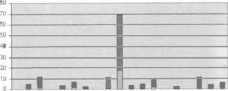
Total Prevalence at depth lOm 83,73%/Location/ Genus-Sp. The dominant coral was bleaching & diseased average is :Acropora, Pocillopora and Montipora. It was occured at loacation Lekuan 2: Symphyllia radians, with Prevalence= 133,33% . Otherwise less Prevalence under 50% was occured at Celah-celah=39,44%, Lekuan 1=33,71% and Fukui=l6,66%.
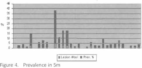
Total Prevalence at depth Sm 30,67%/Location/ Genus-Sp. The dominant coral was bleaching & diseased average is Porites sp and Acropora. High category of prevalence was occurred in Waisai 1=82,13%. Otherwise in Saonek prevalence=S,26%. Is indicate that Sm depth include very good ecosystem ex: water quality, etc.
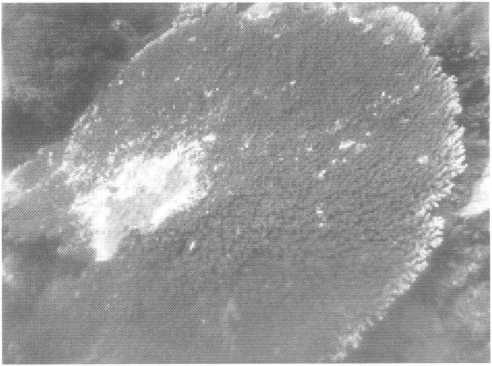
Figure 5. Acropora at Sm depth in Saonek Monde with white band diseased.
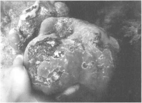
Figure 7. Porites in Saonek Monde at Sm depth with Pigmentation Pesp(lnse (Phn+r r '^-
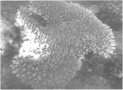
Table. 5. Species Encountered in Sm depth
Figure 6. Acropora at Sm depth with prey gastropoda (Drupe/la corn us).
|
Table. 5. Species Encountered in lOm depth | |||
|
No |
Location |
Coordinates |
Genus/Species |
|
1 |
SaonekMonde (SN) |
unrecord |
Acropora (2x), Hydnopora |
|
2 |
Tanjung Saleo (TS) |
00 ° 26.475' 130 ° 46.222' |
Porites (2x), Montipora |
|
3 |
Waisai 1 (WTC1) |
00 ° 26.019’ 130 ° 49.293' |
Sand |
|
4 |
Waisai2 (WTC2) |
00 ° 25.43 !' 130 ° 50.868' |
Sand |
|
5 |
Saonek (SNK) |
00 ° 28.201' 130 ° 47.281’ |
Acropora (2x), Porites (2x) |
|
Response. | |||
Total Prevalence at lOm depth 23,50%/Location/ Genus-Sp. The dominant coral was bleaching & diseased average is Acropora and Por ites. The area with high prevalence was occured at Tanjung Saleo=61 %. Other-w1'.->e in Saonek Monde=ll,83% (less prevalence). At 1 Om depth the level ofbiodiversity especially coral reef very rare or may even unexist in site: Waisai 1 and 2
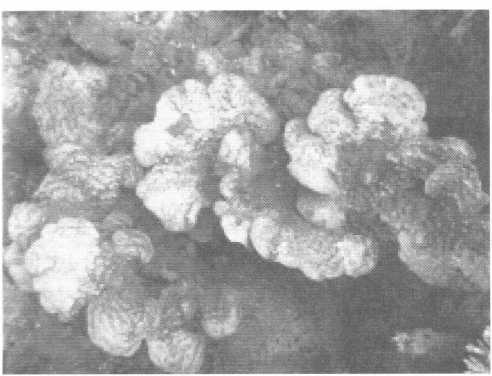
Figure 9. Montipora in Tanjung Saleo at lOm depth with "white syndrome''.
Runduma Island (Wakatobi)
No Location
l Runduma l
2 Runduma2
3 Runduma Anano 1
4 Runduma Anano2
S Runduma Anano3
Coordinates Genus/Species
0)5° 21. 115>, Porites sp, Pocyllopora
124 ° 21.583'
0)5° 2.0). 843' Pocylopora, Porites sp, Acropora
124 ° 20.073'
OS0 17. 396' Porites sp (6x)
124 ° 17.186'
0)5° 17. 789’ Pocylopora sp
124 ° 17. 312'
0)5° 18. 2.38' Porites sp, Lobophyillia, Acropora
124 °17. 930’
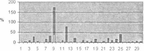
Total Prevalence at Sm depth 23,SS%/Location/ Genus-Sp. The dominant coral was bleaching & diseased average is Porites sp in Runduma Anano 1 with Prevalence = 74,59%
Pre\la Ef"ICe Ble:eching aid Diseased in 10m
Ia Lesion #looI• Pre v. ^ j
I , , 8. Prevalence 1n lOm
Preval eno;a Bl eachi ng and [l seased in 10 m

Figure 12. Prevalence in lOm
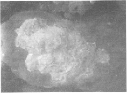
Figure 11. Stoney coral genus Porites at Sm depth with pigmentation response.
Total Prevalence at lOm depth 50,94%/Location/ Genus-Sp. The dominant coral was bleaching e!r diseased aver:1ge is Porites sp. In Runduma 1 with Preva-lence= 185,42%.
Table. 6. Species Encountered in lOm depth
No Location Coordinates
I Runduma l Os0 21.11S^
124° 21.583'
2 Runduma 2 Os0 20. 843'
124° 20.073'
3 Runduma Anano I 05° 17. 396' 124° 17. 186'
4 Runduma Anano 2 05° 17. 789'
124° 17. 312'
S Runduma Anano 3 Os0 18. 238' 124° 17. 930'
Genus/Species
Porites sp (S x)
Porites sp, Pocylopra
Porites sp (4 x), Lobophyllia
Pocylopora sp (2x), Acropora sp, Montipora
Porites sp (2x), Acropora sp (2x)
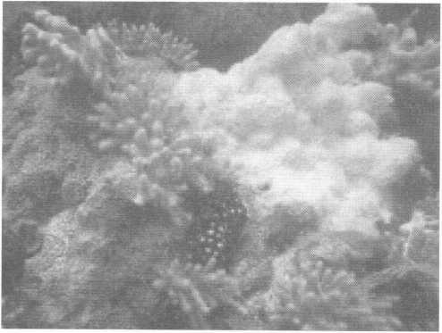
Figure 13. Porites with bleaching pattern. (Foto: E. Ampou)
Ftevaler.::e Bleadiing and Ci seased in 5m·
I a Lesion #/ool • P"'" '1'
Figure 10. Prevalence in Sm
-
1. The high prevalence was occurred at Bunaken National Park especially at lOm depth : 83,73 (poor), low prevalence in Raja Ampat at 1Om depth : 23,50 (good).
-
2. The coral dominant bleaching and diseased is Porites and Acropora, otherwise Pocillopora and Montipora undominant.
SUGGESTION
-
1. The ideal approach to studying coral diseases/ bleaching and their impacts, given sufficient funding and qualified personnel, is a well designed, integrated, multi component survey.
-
2. The result of this research need to be continued in the future such as long term monitoring at the same location to gathering more significant data.
I
ACKNOWLEDGEMENTS
The authors grateful to Balai Riset dan Observasi Kelautan - BR.KP, DKP with DIPA funding, we would like to sincerely thank to Head of BROK for reviewers, comments and suggestions significantly improved the manuscript and Marine Conservation Research Team BROK, TNC-WWF Wakatobi, Conservation International Raja Ampat and Marine Affairs and Fisheries District North Sulawesi, Wakatobi, Raja Ampat for cooperation during collecting data.
REFERENCES
Anonimous. 2007. Siaran Pers : Enam Negara Sepakati Kerjasama Kelola dan Konservasi Segitiga Karang. No 90.PDSI/XII/2007.
Anonimous. 2010. Coral Bleaching. Di unduh dari Wikipedi http://en.wikipedia.org/wiki/Coral_bleaching pada tanggal 6Januari 2010
.Buchheim J., 1998. Coral Reef Bleaching. Marine Biology Learning Center Publications. Di unduh dari http:// www.marinebiology.org/coralbleaching.htm pada tanggal 10 januari 2010.
Buddemeier, RW and Wilkinson, CR. ( 1994) Global Cimate Change and Coral Reefs: Implications for people and reefs. IUCN: Gland (Switzerland), 107 pp.
Coral Disease Handbook, Guidelines forAssesment & Management. 2008. www.gefcoral.org
Dahuri, R. 2003. Keanekaragaman Hayati Laut aset pembangunan berkelanjutan Indonesia. PT Gramedia Pustaka Utama.Jakarta.
Glynn, P.W 1996. Coral reef bleaching: Facts, hypotheses and implications. Global Change Biology 2:495-509.
Kushmaro, A., Y. Loya, M. Fine, and E. Rosenberg. 1996. Bacterial infection and coral bleaching. Nature 380:396.
Hiroya Yamano*, Masayuki Tamura. 2003. Detection limits of coral reef bleaching by satellite remote sensing: Simulation and data analysis. Social and Environmental Systems Division, National Institute for Environmental Studies, Ibaraki,Japan.
Hoegh-Guldberg, 0. (1999) Climate Change, coral bleaching and the future of the world's coral reefs. Greenpeace: Sydney (Australia), 28 pp.
Juliann Krupa. Coral Bleaching and the Affect of Temperature Change on Coral Reef Predator-Prey Interactions. Di unduh dari : http://www.resnet.wm.edu/-jxshix/ math345/juliann-Coral-Bleaching.ppt pada tanggal 12 Januari 2010
Peter J. Mumby,*, William Skirving, Alan E. Strong, John T. Hardy, Ellsworth F. LeDrew, Eric J. Hochberg, Rick P. Stumpf, Laura T. David.Remote sensing ofcoral reefs and their physical environment. Marine Pollution Bulletin48 (2004) 219-228
Peters, E.C. 1997. Diseases of coral reef organisms. In: Birke-land, C. (ed.), Life and Death of Coral Reefs. New York: Chapman & Hall. pp.114-139.
Santavy, D.L. and E.C. Peters. 1997. Microbial pests: Coral disease research in the western Atlantic. Proc. 8th Int. Coral Reef Symp. 1:607-612.
Tyler R. L. Christensen, 2008. Coral bleaching, satellite observations, and coral reef protection. Di unduh dari http://www.eoearth.org/article/Coral_bleaching,_sat: ellite observations,_and_coral reef_ protection pada tanggal 8Januari 2010
The Nature Conservancy. www.coraltrianglecenter.org/. 10 Agustus 2008
http://oceancolor.gsfc.nasa.gov/
http:/ I coralreefwatch.noaa.gov I satellite/virtual-stations/ coral_triangle_virtualstations.html
128
Discussion and feedback