DERMAL CARCINOMA IN DOG AT ZOO CLINIC MAKASSAR
on
Buletin Veteriner Udayana Volume 15 No. 5: 841-846
pISSN: 2085-2495; eISSN: 2477-2712 Oktober 2023
Online pada: http://ojs.unud.ac.id/index.php/buletinvet https://doi.org/10.24843/bulvet.2023.v15.i05.p19
Terakreditasi Nasional Sinta 4, berdasarkan Keputusan Direktur Jenderal Pendidikan Tinggi, Riset, dan Teknologi No. 158/E/KPT/2021
DERMAL CARCINOMA IN DOG AT ZOO CLINIC MAKASSAR
(Dermal Carcinoma pada Anjing di Zoo Klinik Makassar)
1Nurul Sulfi Andini1*, Wa Ode Santa Monica1, Sri Deniaty2, Wahyuni3, I Wayan Nico Fajar Gunawan4
-
1Veterinary Medicine Study Program, Hasanuddin University Perintis Kemerdekaan Km.10 Kota Makassar, Sulawesi Selatan, Indonesia, 90245;
-
2Vet Practicioner in Zoo Clinic, Jl. Metro Tj. Bunga No. 20, Tj. Merdeka, Kec. Tamalate, Kota Makassar, Sulawesi Selatan, Indonesia 90244;
-
3Pathology Laboratory in Balai Besar Veteriner Maros, Jl. DR. Ratulangi, Allepolea, Kec. Lau, Kabupaten Maros, Sulawesi Selatan, Indonesia 90514;
-
4Laboratory of Veterinary Clinical Diagnostic, Clinical Pathology and Radiology. Faculty of Veterinary Medicine, Udayana University. Jl. Raya Sesetan, Gg. Markisa, No. 6 Denpasar
Selatan, Bali, Indonesia, 80225.
*Corresponding author email: nsandini242@gmail.com
Abstract
Dermal carcinoma or skin cancer in dog is a skin cancer consisting of various types of cancer which are included in it. The tumor appears as a solitary, non-encapsulated lump, generally hairless, and occasionally ulcerated, which moves freely. The diagnosis is generally made through microscopic examination of a sample of tumor cells (biopsy). Dermal carcinoma cases are cases that often occur in pets but reports of these cases are still rare especially in Indonesia. The purpose of writing is to describe clinical manifestations, clinical course, and case prognosis. This case report was obtained from the Zoo Clinic Makassar. A dog "Pororo" came with the owner with a temperature of 38.40 C, had an enlarged mass in the medial part of the radius dexter, encapsulated, with an estimated diameter of about 6 cm. Physical examination was carried out, followed by laboratory examination found that the WBC level was very high and anemia. Handling of cases in the form of removal of the tumor through surgery and followed by histopathological examination with Haematoxylin Eosin staining to determine tumor development stage, degree malignancy, prognosis and therapy. The results of histopathological examination stated that the cells found were not uniform and lead to metastasis. The diagnosis of Pororo disease is carcinoma dermatitis but the type is not clear because the primary cells have not been found. The prognosis is infausta, the presence of metastases worsens the prognosis of cases, and it is possible that new tumors will reappear after removal. Handling for similar cases should be done quickly and precisely so that the case prognosis can be better.
Keywords: carcinoma; dermatitis; metastases
Abstrak
Dermal karsinoma atau kanker kulit pada anjing merupakan kanker yang terdiri atas berbagai jenis kanker yang termasuk didalamnya. Tumor muncul sebagai benjolan soliter, tidak berkapsul umumnya tidak berbulu, dan kadang-kadang mengalami ulserasi yang bergerak bebas. Diagnosis umumnya dibuat melalui pemeriksaan mikroskopis dari sampel sel tumor (biopsi). Kasus dermal carcinoma merupakan kasus yang sering terjadi pada hewan kesayangan namun pelaporan mengenai kasus tersebut masih sangat jarang khususnya di Indonesia. Tujuan penulisan untuk mendeskripsikan manifestasi klinis, perjalanan klinis, dan prognosis kasus. Laporan kasus ini diperoleh dari Zoo Klinik Makassar. Seekor anjing “Pororo” datang bersama owner dengan temperature 38,40 C, memiliki massa yang membesar di bagian medial os radius dexter, berkapsul, dengan perkiraan diameter sekitar 6 cm. Pemeriksaan fisik dilakukan, dilanjutkan dengan pemeriksaan laboratorium ditemukan bahwa tingkat WBC sangat tinggi dan anemia. Penanganan kasus berupa pengangkatan tumor melalui operasi dan dilanjutkan dengan pemeriksaan histopatologi dengan pewarnaan Haematoxylin Eosin untuk menetapkan stadium
perkembangan penyakit,derajat keganasan, prognosis dan terapi. Hasil pemeriksaan histopatologi menyatakan sel-sel yang ditemukan sudah tidak seragam dan mengarah ke metastasis. Diagnosis penyakit Pororo adalah karsinoma dermatitis namun dengan type yang belum jelas dikarenakan sudah tidak ditemukannya sel – sel primer. Prognosa kasus infausta, adanya metastasis memperburuk prognosis kasus, dan kemungkinan tumor baru akan muncul kembali pasca pengangkatan. Penanganan untuk kasus serupa sebaiknya dilakukan secara cepat dan tepat sehingga prognosa kasus kemungkinan besar akan lebih baik.
Kata kunci: carcinoma; dermatitis; metastasis
INTRODUCTION
Dermal carcinoma is a skin cancer in dogs generally consisting of melanoma, mast cell tumor/carcinoma, histiocytomas (benign/malignant) and others. Melanoma is a darkly pigmented skin tumor that can be benign (not cancerous) or malignant (cancerous). These tumors or cancers can appear as spots or patches, or raised or flat masses. Most of them have a dark surface (Villalobos, 2019). Histiocytoma are poorly defined skin diseases, all of which are characterized by the proliferation of cells called histiocytes (tissue macrophages). The cause of this disease is unknown. Histiocytoma is a common skin tumor usually seen in younger dogs (less than 3 ½ years) with bad prognosis (Villalobos, 2019; Fulmer, 2007).
The tumor presents as a solitary, raised, generally hairless, and occasionally ulcerated lump that moves freely. Diagnosis is made through microscopic examination of a sample of tumor cells (biopsy). Canine histiocytoma is usually considered a benign tumor. Canine histiocytic neoplasms including skin histiocytoma. These tumors have varied biological behavior, in malignant conditions they often have a poor prognosis (Villalobos, 2019; Fulmer, 2007)
RESEARCH METHODS
History/Anamnesis
A mixed domestic dog, female, aged ± 5 years, there is an enlarged mass on the medial radius. The consistency is hard and has a capsule feel.
Signalement
Pororo, Breed Mix domestic, age ±5 years, has an enlarge mass
Clinical Finding
There is an enlarged mass about 6 cm in diameter in the medial of the right os. The mass is between the skin and muscle fibers. Diagnostic Support
With laboratory examination use complete blood count (CPC) and histopathological examination to determine the type of tumor based on the tissue of origin.
Diagnosis
Carcinoma dermatitis.
Therapy
Tumor removal surgery followed by postoperative observation. Post treatment observation is the surgical wound gradually improving.
RESULTS AND DISCUSSON
Results
The pororo dog has an enlarged mass medial to the os radius dexter, encapsulated, with an estimated diameter of about 6 cm (Figure 1). Preoperative examination was carried out. The preoperation temperature of 38.4 C is normal. The normal temperature for dogs is 37 C-38.4 C (Ngabdussani, 2006). In addition to checking the temperature, a complete blood count (CBC) is also carried out. This blood test is carried out to check the patient's overall health and detect various existing disorders, for example whether there is anemia (decreased hemoglobin level) and infection (increased leukocytes). Based on the CBC examination results on Pororo, it
was found that the levels of WBC (leukocytes), neutrophils, eosinophils, and basophils increased. RBC, HGB (Hemoglobin), HCT, MCV decreased while MCH and MCHC increased (Table 1). Increased WBC indicates an infection, this is also supported by the high number of neutrophils, eosinophils and basophils.
The results of Pororo's blood examination, Pororo had hyperchromic microcytic anemia which was characterized by a decrease in MCV while MCH and MCHC increased. Anemia can be caused by one of three mechanisms, namely blood loss (eg bleeding), increased destruction of erythrocytes (by the process of hemolysis), and decreased production of erythrocytes (Price, 2006).
Tumor removal is done by excising all of the tumor tissue and a small amount of surrounding healthy tissue (Figure 2). The skin is cut around the tumor, after the skin is exposed a hard tumor appears. The tumor is removed after the small blood vessels around it are tied. After removal of the tumor, the incision wound was sutured using simple interrupted sutures (Figure 2). Then given betadine and nebacetyn powder. The tumor mass that has been removed is then subjected to histopathological examination. The results of histological examination showed that there was severe inflammation (Figure 3), which in turn also showed a collection of cells that were not uniform and led to the occurrence of metastases (Figure 4). The results of histopathological examination showed that the tumor that formed on the medial os.radius dexter of the Pororo dog was a malignant (cancerous) tumor. Primary cells which are the initial or basic diagnosis for determining the type of cancer are not mentioned because the cells formed are in the form of cells that are not uniform in large numbers.
Discussion
Dermal carcinoma is often cases in pets (Widyarini, 2022). But the report of this cases is still rare especially in Indonesia. The diagnosis of the main tumor was
carried out by classical histopathological examination (Widyarini, 2022). In this case, we still cannot identify what kind of the skin cancer that Pororo had. The results of histopathological examination showed that the tumor that formed on the medial os radius dexter of the Pororo was a malignant (cancerous) tumor. Primary cells which are the initial or basic diagnosis for determining the type of cancer are not mentioned because the cells formed are not uniform in large numbers. In general, skin tumors in dogs from mesenchymal tissue, and another 45% derived from the epithelium. Tumor the most common mesenchyme in dogs. These are histiocytoma, lipoma, and tissue cell tumors fat, and fibrosarcoma (Widyarini,2022).
There is a possibility that this case leads to fibrosarcoma, this is in accordance with the opinion of Widyarini (2022), microscopic appearance of fibrosarcoma in skin in the form of tumor cell size and shape tumor cell nuclei vary. Shaped tumor cells spindle shape tumor with pattern interwoven or herring bone pattern with density high core. The same thing was also stated by Hendrick (2016) fibrosarcoma can be circumscribed or infiltrative, small or extremely large and disfiguring. Capsules are usually not seen. However, for the metastatic case, still hard to define the histogenesis.
Metastasis is defined as the spread of cancer cells originating from the primary tumor to different tissues and organs. Metastasis, which is the most dangerous stage of cancer development, complicates disease treatment and can cause patient death (Topcul, 2012). Metastatic spread of neoplasms to the skin is uncommon. The three common routes of spread are either by lymphatics, by blood vessels, or by implantation at the time of surgery. Immunohistochemistry and the appropriate history of a primary tumor at a distant site are helpful in establishing the diagnosis of a metastatic tumor (Goldschmidt, 2016).
The clinical implications of increased neutrophils are bacterial infections,
metabolic disturbances, bleeding and myeloproliferative disorders. If the histamine concentration is increased, the basophil level is usually high. The clinical implications of eosinophilia are the body's response to neoplasms, Addison's disease, allergic reactions, collagen vascular disease or parasitic infections. Basophils are associated with granulocytic leukemia and basophilic myeloid metaplasia and allergic reactions. For cases of eosinophilia and basophilia, this is associated with parasitic diseases related to systemic circulation such as heartworms, tick infestations and dermatitis (Wilyanto, 2018)
Decreased Hb values can occur in anemia (especially anemia due to iron deficiency), cirrhosis, hyperthyroidism, bleeding, increased fluid intake and pregnancy. HCT (Hematocrit) shows the percentage of red blood cells to the total blood volume. Decreased Hct values are an indicator of anemia (due to various causes), hemolytic reactions, leukemia, cirrhosis, large blood loss and hyperthyroidism. Anemia can be caused by one of three mechanisms, namely blood loss (eg. bleeding), increased destruction of erythrocytes (by the process of hemolysis), and decreased production of erythrocytes (Price, 2006).
Premedication and sedation were carried out using ketamine and xylazine each as much as 1 ml intramuscularly. After the dog fell asleep, the hair around the tumor was shaved and cleaned with 70% alcohol and liquid betadine. Then given RL infusion fluids.
Postoperative treatment with administration of drugs in the form of Betamox LA and Dexametahosone. Postoperative care was carried out by cleaning the cage and wound conditions with povidone iodine. This is in accordance with Kumar (2015) antiseptic dressing of the suture line with povidone iodine is recommended for postoperative lipoma tumors. The healing process was relatively fast, on the third day after surgery, the
wound began to dry. Day 7 after surgery the patient was active again and went home.
CONCLUSION AND SUGGESTIONS
Conclusion
Pororo case was diagnosed as dermal carcinoma or skin cancer, but the type was not clear. This is because the primary cells to determine the type of cancer are not found due to metastases. The process of handling cases of dermal carcinoma can be done by surgery followed by giving antibiotics to postoperative patients. But do not rule out the cancer mass can reappear.
Suggestion
Treatment of cancer in particular must be done quickly and precisely. In the case of the Pororo dog, the diagnosis of cancer was relatively late because the cells had metastasized.
ACKNOWLEDGEMENT
We thank to Zoo Clinic Makassar for allow us to follow and write this case.
REFERENCES
Fulmer KA, Mauldin GE. 2007. Canine histiocytic neoplasia: An overview. Can. Vet. J. 48(10): 1041–1050.
Goldschmidt MH, Goldschmidt KH. 2016.
Epithelial and melanocytic tumors of the skin. Tumors in Domestic Animals, 5th Ed. Wiley Blackwell. Pennsylvania. Pp. 88–141.
Hendrick MJ. 2016. Mesenchymal tumors of the skin and soft tissues. Tumors in Domestic Animals. 5th Ed. WileyBlackwell. Pennsylvania. Pp. 142-175.
Kumar KMN, Laksmi DP, Veena P, Reddy KS. 2015. Surgical management of lipoma in a dog. Int. J. Sci. Environ. Technol. 4(5): 1301-1304.
Ngabdussani I. 2006. Nilai Fisiologis kardiovaskuler, respirasi, suhu tubuh anjing dewasa dan anak. Skripsi. Bogor, Institut Pertanian Bogor.
Price SA, Wilson LM. 2006. Patofisiologi: konsep klinis proses proses penyakit, 6th Ed. Jakarta: EGC.
Topcul M, Cetin I. 2012. Cancer stem cells in oncology. J. BUON. 17(4): 644-648.
Villalobos A. 2019. Tumours skin in dogs. Merck and the Merck Veterinary Manual.
https://www.merckvetmanual.com/dog -owners/skin-disorders-of-dogs/tumors-of-the-skin-in-dogs.
Widyarini S, Sugiyono, Kristiangingrum YK, Sutrisno B. 2022. Studi histopatologis tumor pada hewan kesayangan dan hewan liar. J. Sains Vet. 40(1): 73-84.
Wilyanto I. 2018. Interpretasi hitung darah lengkap. Inl Veterinary Service Surabaya.
https://slideplayer.info/slide/11952037/
Table 1. Blood Test Result for Pororo
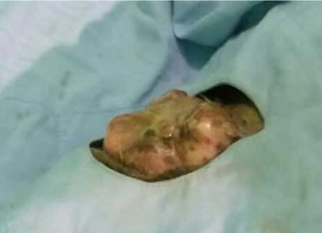
Figure 1. Enlarges mass in skin of Pororo
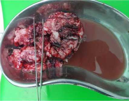
Figure 2. Removed tumour tissue from
Pororo’s skin
|
Type |
Score |
Interpretation |
|
WBC |
32,17 +109 /l |
High |
|
NEU |
25.98 +109 /l |
High |
|
EOS |
1,32 +109 /l |
High |
|
BAS |
0,48+109 /l |
High |
|
RBC |
4,02- 10 12 |
Low |
|
MCV |
55-fl |
Low |
|
MCH |
27,3 + pg |
High |
|
MCHC |
49,4 +g/dl |
High |
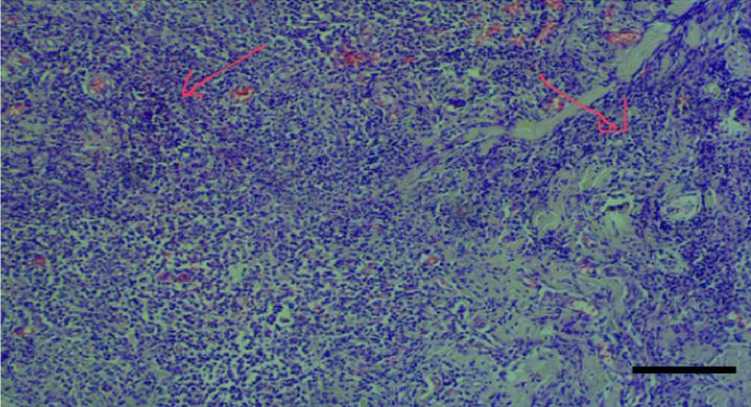
Figure 3. Histopathology samples of Pororo’s has intense inflammation. Inflammation was described by red arrows. HE, 40x
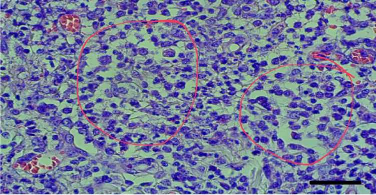
Figure 4. Histopathology of Pororo’s has non-uniform cells leads to metastasis, eas describe by red circle. HE, 40x
846
Discussion and feedback