POTENTIAL EFFECT OF MACRO ALGA Caulerpa sp. AND Gracilaria sp. EXTRACT LOWERING MALONDIALDEHYDE LEVEL OF WISTAR RATS FED HIGH CHOLESTEROL DIET
on
INTERNATIONAL JOURNAL OF BIOSCIENCES AND BIOTECHNOLOGY • Vol. 5 No. 1 • September 2017 ISSN: 2303-3371 https://doi.org/10.24843/IJBB.2017.v05.i01.p06
POTENTIAL EFFECT OF MACRO ALGA Caulerpa sp. AND Gracilaria sp. EXTRACT LOWERING MALONDIALDEHYDE LEVEL OF WISTAR RATS
FED HIGH CHOLESTEROL DIET
K. Srie Marhaeni Julyasih1* and I G. P. Wirawan2
-
1Faculty of Agriculture, UPN”Veteran” East Java
2
-
2Faculty of Agriculture, University of Udayana, Bali-Indonesia *Corresponding author: smjulyasih@gmail.com
ABSTRACT
Seaweed has potential nutrient content such as carotenoids, vitamins, fatty acids, carbohydrates, minerals, and other essential substances. Carotenoids have important biological functions as an antioxidant, and immunostimulatory which can prevent the disease, anti-inflammatory, anti-stress, anti-aging, and protect the skin from the harmful effects of ultraviolet radiation. Seaweed generally consumed as a vegetable by people in Bali, known as the local name Bulung Boni (Caulerpa spp.) and Bulung Sangu (Gracilaria spp.).. So far there has been no report or results of research on the effects of extract ethanol of Bulung Boni (Caulerpa sp.) and Bulung Sangu (Gracilaria sp.) as an antioxidant that can prevent lipid peroxidation which can be seen in decreased level of MDA in liver tissue or blood plasma. Therefore it is necessary to determine of plasmaMDA level of Wistar rat after fed high cholesterol diet treated with extract ethanol of Caulerpa sp. and Gracillaria sp. This experimental study used Completely Randomized Design. Research using total of 24 Wistar rats divided into six sample groups of equal size, all fed with a diet high in cholesterol especially in negative control. The study consisted of negative control group (standard diet), positive control group (high cholesterol diet), high-cholesterol diet with Caulerpa sp. extract dose of 20 mg and 60 mg/100 g, high cholesterol diet with Gracilaria sp. extract dose of 20 mg and 60 mg/100 g body weight rat per day.The study resulted that rats fed high cholesterol diet with treated extract ethanol Caulerpa sp. and Gracilaria sp. with a dose of 20 mg and 60 mg per 100 g body weight rat / day had plasma MDA level significantly lower (p <0.05) compared with rats fed high cholesterol diet without treated with extract of Caulerpa sp. and Gracilaria sp.
Keywords: Caulerpa sp., Gracilaria sp., MDA, and seaweeds
INTRODUCTION
In some countries such as Japan, Korea, China, Vietnam, Indonesia, Peru, Scandinavia, Scotland, and Philippines, seaweed has been used as a source of food, medicine, and raw material for
various types of industries. Seaweed has potential nutrient content such as carotenoids, vitamins, fatty acids, carbohydrates, minerals, and other essential substances. Carotenoids have important biological functions as an
antioxidant, and immunostimulatory which can prevent the disease, antiinflammatory, anti-stress, anti-aging, and protect the skin from the harmful effects of ultraviolet radiation (Kato et al., 2004; El-Baky et al., 2007). Carotenoids are antioxidant that are potential in protecting against membrane lipid peroxidation (Siems et al., 2002). Carotenoids are derived from natural sources more safe than synthetic carotenoids (Allan, 2006).
Antioxidant can protect the body from free radical attack and reduce its negative impact. Free radicals are necessary for the survival of several physiological processes in the body, especially for electrons transport, but the excessive free radical can harm the body because it can damage macromolecules such as protein in cells, and DNA (deoxyribo nucleic acid). Macromolecular damage can improve cell death (Haliwell, 2002). Malondialdehyde (MDA) can be used as
-
K. Srie Marhaeni Julyasih and I Gede Putu Wirawan an indicator of lipid peroxidation by free radical (Aksoy et al., 2003).
Normally the body has a systematic strategy to counteract the free radical or to accelerate degradation of these compounds. This system can be divided into two groups: preventive defense systems such as the enzyme superoxide dismutase (SOD), catalase, glutathione peroxidase, and defense systems through the termination of radical reaction such as α-tocopherol, vitamin C, and carotenoids (Kumalaningsih, 2007). Antioxidant compounds derived from plants such as vitamin C, vitamin E, carotenoids, phenolic groups, especially polyphenols, and flavonoids have potential effect to reduce the risk of degenerative diseases (Amrun et al., 2007).
In addition besides effects of oxidants, cholesterol also affects the development of degenerative diseases. The development of people live style that consume more fatty foods, especially of saturated fatty acid intake tend to
cholesterol to be higher than the level of need. Intake of foods with high cholesterol content can increase cholesterol levels in the blood. This condition called hypercholesterolemia. One of the major atherosclerosis risk factors are dyslipidemia, and the prevalence of dyslipidemia in Indonesia has increased (Anwar, 2006).
The treatment of patients with hypercholesterolemia require a long time, and high costs, the research continues to be developed to obtain a more effective drug with a cheaper price, and reduce side effects. Various studies of antioxidants also still needs to be done considering the huge benefits for health. Natural ingredients from the sea needs to be explored because the content of its bioactive especially antioxidants has not been thoroughly explored. As one effort to optimize the utilization of marine natural products of Indonesia, it is necessary to do research on seaweed.
In Bali there are several types of seaweed that is generally consumed by people known as the local name Bulung Boni (Caulerpa spp.) and Bulung Sangu (Gracilaria spp.). Seaweed is often consumed as a vegetable or snack and have been consumed hereditary. So far there has been no report or results of research on the effects of Bulung Boni and Bulung Sangu extract as an antioxidant that can prevent lipid peroxidation which can be seen in decreased level of MDA in liver tissue or blood plasmaThe results can provide information to the public about Bulung Boni and Bulung Sangu benefits can be used as natural antioxidants to prevent the effects of free radicals which is one risk factor for the development of degenerative diseases.
MATERIALS AND METHODS
The study was conducted with Experimental research measurement the levels of plasma malondialdehyde Study using completely randomized design
(Murdiyanto, 2008). The study consisted of negative control group (standard diet), positive control group (high cholesterol diet), rats fed high-cholesterol diet with Bulung Boni extract dose of 20 mg and 60 mg/100 g, rats fed high cholesterol diet with Bulung Sangu extract dose of 20 mg and 60 mg/100 g body weight rat per day.
Seaweeds was collected from Serangan Beach Bali. As for the further analysis carried out at the Laboratory of Healthy Plant, UPN "Veteran" Jawa Timur, Agricultural Biotechnology Laboratory Udayana University and Laboratory of Pharmacology Faculty of Medicine Udayana University.
Preparation of seaweeds extract
Seaweed is dried and then crushed in a blender , mixture with ethanol filtered
by filter paper Whatman 42, evaporated with vacuum evaprator to result crude extract..
Extract of seaweed treated in rats
In one cage was placed as many as four rats that had previously adapted for one week in the laboratory. Standard diet, cholesterol diet, and beverages rats administered daily ad libitum. Seaweed extract was administered orally by zonde with dose 20 mg and 60 mg/100 g body weight rat/day according to treatment.
Measurement of MDA Level
After 30 days treatment, rats was fasted for 18 hours. Bloods sample was taken through the sinus orbitalis as much as 2 cc. Measurement of MDA levels using Thiobarbituric Acid Reactive Substances (TBARS). (Rahayu, 2005).
Data analysis
Statistical analysis of data using SPSS for windows . To determine the effect of treatment, the data were analyzed by analysis of variance at a significance level of 5 %. If the F-test
showed a significant difference then , further treatments were tested with LSD at the 5 % significance level .
RESULTS AND DISCUSSION
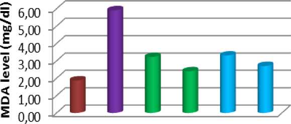
The lowest plasma MDA level found in the negative control is 1.88 ± 0.22 mg / dl, then Caulerpa 60 mg is
2.41 ± 0.10 mg / dl, Gracilaria 60mg is 2.71 ± 0.17 mg /dl, Caulerpa 20 mg is 3.22 ± 0.47 mg / dl, Gracilaria 20 mg is 3.31 ± 0.19 mg /dl, and the highest in the positive control with plasma MDA level of 5.91 ± 0.22 mg /dl (Fig. 1).
Plasma MDA Level


-
■ negative control
-
■ positive control ICauIerpa 20 mg
-
■ Caulerpa 60 mg BGraciIaria 20mg BGraciIaria 60 mg
Fig. 1. Plasma MDA Level Wistar Rat in Negative Control, Positive Control, Caulerpa 20 mg, Caulerpa 60 mg, Gracilaria 20 mg, and Gracilaria 60 mg.
Analysis of variance of plasma MDA level Wistar rat treated high-cholesterol diet with Bulung Boni
(Caulerpa sp.) and Bulung Sangu
(Gracilaria sp) extract showed
significantly different (p <0.05) in various
treatments. To determine the effect of each treatment on plasma MDA level performed with multiple comparation test. Plasma MDA level in Caulerpa 60m significantly lower compared with Caulerpa 20 mg, Gracilaria 20 mg, and positive control, but did not differ significantly with Gracilaria 60 mg. Plasma MDA level in negative control significantly lower compared with other treatment.
Provision of high-cholesterol diet resulted in plasma MDA levels were significantly higher than other treatments, this is likely due to the oxidative damage to the unsaturated fat. The fatty acid chain polyunsaturated phospholipid membrane layer is attacked by hydroxyl radicals cause lipid peroxidation. Hoarding on the membrane lipid hydroperoxide will cause interference with the function of cells. Lipid peroxides can then be turned into toxic compounds, namely aldehydes, MDA, and hydroxy nonenal. The
-
K. Srie Marhaeni Julyasih and I Gede Putu Wirawan concentration of MDA in the biological material can be used as an indicator of damage okdidatif in unsaturated
fats, as
well as an indicator of the presence of free radicals.
MDA analysis is an analysis of free radicals indirectly and easily determine the number of free radicals are formed. Analysis of free radicals directly is very difficult to do because the radicals are very unstable. Measurements can be made by reacting MDA TBARS (Hurry et al.,
-
2002). Provision of high-cholesterol diet and extracts Caulerpa 20 mg or Gracilaria per 100 g bw rat per day, can reduce levels of MDA plasma, thus significantly lower than rats that were only given high cholesterol diet. The extract Caulerpa sp and Gracilaria sp. with a higher dose of 60 mg per 100 g bw rat resulted in an average of plasma MDA levels were significantly lower compared with to Caulerpa 20 mg,
Gracilaria 20 mg, and positive control.. Increasing doses of the extract resulted in more active ingredient in the extract, so the ability to reduce levels of plasma MDA higher. Carotenoids are antioxidants and can capture free radicals. According Ardiansyah (2007), the antioxidant defenses naturally in LDL cholesterol by an amount sufficient to protect LDL from oxidation. Beta carotene is a fairly strong antioxidants which theoretically also can protect LDL oxidation.
CONCLUSIONS
Plasma MDA level of Wistar rat fed high cholesterol diet treated with Caulerpa sp. and Gracilaria sp. extract with doses of 20 mg and 60 mg significantly lower compared with Wistar rat fed high-cholesterol diet without treated Caulerpa sp. and Gracilaria sp. extract.The lowest plasma MDA level found in the negative control is 1.88 ± 0.22 mg / dl, then Caulerpa 60 mg is
-
2.41 ± 0.10 mg / dl, Gracilaria 60mg is
-
2.71 ± 0.17 mg /dl, Caulerpa 20 mg is
-
3.22 ± 0.47 mg / dl, Gracilaria 20 mg is 3.31 ± 0.19 mg /dl, and the highest in the positive control with plasma MDA level of 5.91 ± 0.22 mg /dl.
REFFERENCES
Adam, J. M. F. (2005). Meningkatkan Kolesterol HDL, Paradigma Baru Penatalaksanaan Dislipidemi. J.Med Nus, 26(3): 200-204.
Aksoy, H., Koruk, M., & Akcay, F.
(2003). The Relationship between Serum Malondialdehyde and Ceruloplasmin in Chronic Liver Disease. Turkish Jurnal of Biochemistry, 28(2): 32-34.
Alan, M. (2006). Carotenoids and Other Pigments as Natural Colorants. Pure Appl.Chem, 78 (8): 1477-1491
Anwar, T.B. (2004). Dislipidemia Sebagai Faktor Resiko Penyakit Jantung Koroner. e-USU Repository
Anonim. (2008). Algal Pigments. Available at:
URL:http://www.clarku.edu/faculty/ Robertson/laboratory20%methods/p igments.html. 5 September 2010.
Ardiansyah. (2007). Antioksidan dan Peranannya Bagi Kesehatan. Available from:URL:
http://www.iptek.net. 23 Januari 2007.
Burtin, P. (2002). Nutritional Value of Seaweeds. Electronic Journal of Enviromental, Agricultural and Food Chemistry, ISSN: 1579-4377.
Colpo, A. (2005). LDL Cholesterol: Bad Cholesterol, or Bad Science. Journal of American Physicians and Surgeons, 10 (3): 83-89
Crowe, K. (2005). Plant Pigment Chemistry: Pigment Extraction and Analysis using Thin Layer Chromatography. University of Marine.
El-Baky, H. H., El-Baz, F. K., & El-Baroty, G.S. (2007). Production of Carotenoids from Marine
Microalgae and its Evaluation as Safe Food Colorant and Lowering Cholesterol Agents. American Eurasian J.Agric. Sci, 2(6): 792800.
Fuhrman, B., Elis, A., & Aviran, M.
(2002). Hyphocholesterolemic
Effect of Lycopene and β carotene is Related to Support of Cholesterol Synthesis and Augmentation of LDL Receptor Activity in Macrophages. Biochemical and Biophysical Research Comunicated. 232(3): 658-662.
Haliwell, B. (2002). Food Derived
Antioxidants: How to Evaluate
Their Importance in Food and in
Vivo. Hand book of Antioxidants.
Second Edition. Revised and
Expandee Edited by Erique Cadences Lester Packer. University of Southern California School of Pharmacy. Los Angeles California. p 1-33.
Hangbao, M., Young, J., & Shen, C.
(2008). HMG-CoA Reductase (3 hydroxy-3 methyl glutaryl-CoA Reductase) (HMGR). Journal of American Science. 4 (3). ISSN 1541003.Availablefrom: URL:http://www.americanscience.o rg
Jae, K.W. (2008). Kolesterol. Yayasan Jantung Indonesia. Available from: URL:http://www.heartinfo.org. 17 September 2008.
Kagami, S. I., Kanari, H., Suto, A., Fujiwara, M., Ikeda, K., Hiroshe, K., Hikowatanabe, N., Iwanoto, I., & Nakajima, H. (2008). HMG-CoA reductase Inhibitor Simvastatin Inhibit Proinflamatory Cytokine
Production from Marine Mast Cells. Int.Arch Allergy Immunol, 146(1): 61-6
Kato, M., Ikona, Y., Matsumoto, H., Sugiura, M., Hyodo, H., & Yano, M. (2004). Accumulation of Carotenoids and Expression of
Carotenoids Biosynthetic Genes
During Maturation in Citrus Fruit. Plant Physiol February, 134 (2):
824-837. Liang Song, B., Javitt, N. B., & Boyd, R. A. D. (2005). Insig Mediated Degradation of HMG-CoA reductase Stimulated by lanosterol, an Intermediate in The Synthesis of Cholesterol. Cell Metabolism, 1: 179-189
Murdiyanto, B. (2008). Rancangan Percobaan. Available from URL: http://www.ikanlaut.tripod.com. 7 Juli 2009.
Myers, S. (2005). The Carotenoids Palette. An Array of Colors, Researched Health Benefits and Formulation Challengers Highlight the Future of Carotenoids. Available from: URL:
http://www.naturalproductsinsider.c om. 23 Juni 2007.
Pengembangan dan Pemanfaatan Obat
Bahan Alam. (1991). Pedoman
Pengujian dan Pengembangan
Fitofarmaka. Penapisan
Farmakologi, Pengujian
Fitokimia, dan Pengujian Klinik. Kelompok Kerja Ilmiah. Jakarta: Yayasan Pengembangan Obat Bahan Alam Phyto Medica.
Rahayu, T. (2005). Kadar Kolesterol Darah Tikus Putih (Rattus
norvegicus L) Setelah Pemberian Cairan Kombucha Per-Oral. Jurnal Penelitian Sains & Teknologi, 6(2): 85 – 100.
Siems, W.G., Sommerburg, O., &
Frederick, J. G. M. (2002). Hand book of Antioxidants. Second Edition. Revised and Expandeed
Edited by Erique Cadences Lester Packer. University of Southern
California School of Pharmacy. Los Angeles California. p 235-245
Suharto., Girisuta, B., & Miryanti, A.
(2004). Perekayasaan Metodologi Penelitian. Penerbit Andi
Yogyakarta. 218p
ASIA OCEANIA BIOSCIENCE AND BIOTECHNOLOGY • 79
Discussion and feedback