MOLECULAR IDENTIFICATION OF BLASTOCYSTIS SP. IN LONG-TAILED MACAQUE AT ALAS PURWO PARK
on
Buletin Veteriner Udayana Volume 15 No. 5: 732-736
pISSN: 2085-2495; eISSN: 2477-2712 Oktober 2023
Online pada: http://ojs.unud.ac.id/index.php/buletinvet https://doi.org/10.24843/bulvet.2023.v15.i05.p05
Terakreditasi Nasional Sinta 4, berdasarkan Keputusan Direktur Jenderal
Pendidikan Tinggi, Riset, dan Teknologi No. 158/E/KPT/2021
MOLECULAR IDENTIFICATION OF BLASTOCYSTIS SP. IN LONG-TAILED MACAQUE AT ALAS PURWO PARK
(Identifikasi Molekuler Blastocystis sp. pada Monyet Ekor panjang di Taman Nasional Alas Purwo)
Edward Yonas Kristijanto1*, Wiwik Misaco Yuniarti2, Mufasirin2, Poedji Hastutiek2, Endang Suprihati2, Boedi Setiawan2, Puput Ade Wahyuningtyas3, Doohan Mahendra4
-
1Master Student, Faculty of Veterinary Medicine, Airlangga University, Surabaya, Indonesia;
-
2Faculty of Veterinary Medicine, Airlangga University, Surabaya, Indonesia;
-
3Institute of Tropical Disease, Airlangga University, Surabaya, Indonesia;
-
4Balai Karantina Pertanian Kelas II Palu, Indonesia.
-
*Corresponding author email: yonaskristijanto2711@gmail.com
Abstract
Zoonotic diseases can be transmitted through close interactions between long-tailed monkeys. Blastocystis sp. is one of the parasites that can attack mammals and is most commonly found in the intestinal tract. This study aims to analyze the presence of infection and the phylogenetic tree of Blastocystis sp. in long-tailed monkeys in Alas Purwo National Park, Banyuwangi, East Java. Identification of Blastocystis sp. in this study using morphological and molecular methods. A total of 100 stools were examined microscopically using the floating method, showing that 61 samples were positive, followed by a PCR test with a target of 600bp. PCR results obtained three positive samples followed by squencing. Sequences processed in BLAST isolate samples TNAP2 and TNAP9 having homology with Blastocystis sp. Subtype 3 was 98-99%, while the TNAP7 isolate had a lower homology level of 78-79% and the level of phylogenetic analysis of the TNAP2 and TNAP9 isolates was related to Blastocystis sp. from the Philippines (KY610153.1) and Egypt (OP942294.1) and the TNAP7 isolate is related to Blastocystis sp. from Thailand (MH197670.1, MH197668.1). Isolate Blastocystis sp. from Alas Purwo National Park has high homology analysis results to Blastocystis type hominis from Rep. Czech and Chinese by 80-99%, it is possible to have a connection with the zoonotic problem.
Keywords: Blastocystis sp.; long-tailed monkeys; Alas Purwo National Park; zoonotic diseases.
Abstrak
Penyakit zoonosis dapat ditularkan melalui Interaksi yang dekat antara monyet ekor panjang. Blastocystis sp. merupakan salah satu parasit yang dapat menyerang mamalia dan paling banyak ditemukan di saluran usus. Penelitian ini bertujuan untuk menganalisis adanya infeksi dan pohon filogenetik dari Blastocystis sp. pada monyet ekor panjang di Taman Nasional Alas Purwo, Banyuwangi, Jawa Timur. Identifikasi Blastocystis sp. pada penelitian ini menggunakan metode morfologis dan molekuler. Sebanyak 100 feses dilakukan pemeriksaan secara mikroskopis dengan metode apung menunjukkan bahwa ada 61 sampel yang positif, dilanjutkan dengan uji PCR dengan target 600bp. Hasil PCR didapatkan tiga sampel positif dilanjutkan dengan squencing. Hasil sekuens diproses dalam BLAST isolate sampel TNAP2 dan TNAP9 memiliki tingkat homologi dengan Blastocystis sp. Subtype 3 sebesar 98-99% sedangan isolate sampel TNAP7 memiliki tingkat homologi yang lebih rendah yaitu 78-79% dan tingkat analisis filogenetik isolat TNAP2 dan TNAP9 memiliki kekerabatan dengan isolat Blastocystis sp. dari Filipina ( KY610153.1) dan Mesir (OP942294.1) serta isolat TNAP7 memiliki kekerabatan dengan isolat Blastocystis sp. dari Thailand (MH197670.1, MH197668.1). Isolat Blastocystis sp. asal Taman Nasional Alas Purwo mempunyai hasil analisis homologi yang tinggi terhadap Blastocystis type hominis asal Rep. Ceko dan China sebesar 80-99%, hal itu memungkinkan mempunyai keterkaitan dalam masalah zoonosis.
Kata kunci: Blastocystis sp.; monyet ekor panjang; Taman Nasional Alas Purwo; penyakit zoonosis.
INTRODUCTION
Blastocystis is a parasite that can attack mammals and is most commonly found in the intestinal tract in various vertebrae including humans (Zhu et al., 2017). Transmission of Blastocystis sp through direct contact with infected animals is a zoonotic risk that can occur (Osman et al., 2015). The interaction between visitors and long-tailed monkeys can cause changes in behavior to become aggressive. This causes the risk of disease transmission between long-tailed monkeys and humans to increase (Friishansen et al., 2015). In developing countries the prevalence distribution of blastocystis infection is up to 100%, while in developed countries it is up to 56% (El Safadi et al., 2014; Scanlan et al., 2014). Blastocystis sp. found in children and monkeys in an area in Khatmandu India (Yoshikawa et al. (2009). Blastocystis sp. identified based on analysis of the small subunit ribosomal RNA (SSU rRNA) gene has 17 subtypes (Alfellani et al., 2013). In research on Blastocystis sp., there are similar subtypes between humans and Non-Human Primates (NHP), ST1, ST2, ST3, and ST4 (Yoshikawa et al., 2009; Alfellani et al., 2013; Ramírez et al., 2016) In determining subtypes and phylogenetic trees to analyze the presence of Blastocystis sp. infection in long-tailed macaques in Alas Purwo National Park, Banyuwangi, East Java, it is necessary to carry out morphological and molecular examinations.
RESEARCH METHODS
Sample Collection
100 samples of long-tailed monkey feces in Alas Purwo National Park, Banyuwangi, East Java were taken 5 grams and then collected and put in pots. sample containing 2% potassium dichromate. Each pot was given a sample code, then put in an ice box and then stored at -4OC until DNA extraction was carried out.
Sample Inspection
Microscopic Examination of
Blastocystis sp. Stool samples were examined at the Laboratory of the Institute of Tropical Diseases, Airlangga University, Surabaya. Samples were examined to observe each phase of Blastocystis namely vacuolar, granular and cyst. Identification for this protozoa using native and floating methods and observed using a microscope with a magnification of 400 and 1000 times. Microscopic identification of Blastocystis is based on vacuolar and granular forms. The vacuolar form has two nuclei located at opposite poles, while the granular form shows small granules that fill the cytoplasm (Arpitha et al., 2018).
Molecular Examination by PCR
Stool samples that have been confirmed through morphological examination are then followed by microscopic examination. There were three samples which were continued at the stage using the PCR method. A sample of 100 µl was put in a 1.5 ml microtube and DNA was extracted using the QIAamp DNA stool mini kit (QIAGEN, Hilden, Germany) according to the protocol in the kit. Furthermore, the samples were stored at -20°C until further examination. This research was conducted at the Institute of Tropical Disease (ITD) Airlangga University, Surabaya. PCR amplification used the primers BhRDr
(GAGCTTTTTAACTGCA-ACAACG) and RD5 (ATCTGGTTGATC-
CTGCCAGT) (Kurniawati et al., 2020). Initial denaturation was carried out at 94°C for 5 minutes, followed by 35 cycles of denaturation at 94°C for 1 minute, annealing at 56°C for 1 minute and
elongation at 72°C for 1 minute, and
additional elongation for 5 minutes. The PCR product was then read on 2% agarose gel electrophoresis and showed the band at the 600 bp position using Ultraviolet light (Mahendra et al., 2020). Samples that have been confirmed through morphological
and molecular examinations, samples that are positive will continue sequencing.
Data analysis
Data from the sequencing results from the samples obtained will be processed in the BLAST NCBI Genbank database and forwarded using a phylogenetic tree with the genetyx ver.10 application.
RESULTS AND DISCUSSION
Results of Morphological Examination of Blastocystis sp
Microscopic morphological analysis of 100 stool samples examined
microscopically showed that 61 samples (61%) were positive for Blastocystis sp. The results of microscopic examination using the floating method showed that the parasite was morphologically similar to Blastocystis sp. (Figure 1). According to Ahmed and Karanis (2018) the morphological form of Blastocystis sp has characteristics, namely the nucleus is located on the edge of the cytoplasm, there is sizable cytoplasm inside the cell, has 1 central vacuole or there is a vacuole in the middle (vacuolar) (Figure 1 A), No there are vacuoles or vacuoles are replaced by granules (granular) (Figure 1 B) and there is no central vacuole (cyst) (Figure 1 C).
Polymerase Chain Reaction (PCR) Examination Results
PCR results with primer Blastocystis sp. BhRDr (5'-GAG CTT TTT AAC TCC AAC AAA CG-3') and RD5 (5'-ATC TGG TTG ATC CTG CCA GT-3') (Kurniawati et al., 2020) which produced a band of 600 bp in amplification. The results of PCR product visualization using electrophoresis can be seen in Figure 2.
PCR results obtained three positive samples followed by squencing. RD5 is a primer that amplifies eukaryotic species widely and BhRDr is a primer with high Blastocystis specificity. False positives can occur, for that confirmation through sequencing is done to confirm the results are positive (Stensvold et al., 2013). Sequences processed in BLAST isolate
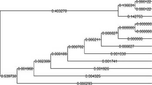
samples TNAP2 and TNAP9 having homology with Blastocystis sp. Subtype 3 was 98-99%, while the TNAP7 isolate had a lower homology level of 78-79% and the level of phylogenetic analysis of the TNAP2 and TNAP9 isolates was related to Blastocystis sp. from the Philippines (KY610153.1) and Egypt (OP942294.1) and the TNAP7 isolate is related to Blastocystis sp. from Thailand (MH197670.1, MH197668.1). Subtypes 1 and 3 are subtypes that are commonly found in humans and non-human primates (NHP) in the world (Ramírez et al., 2016). Isolate Blastocystis sp. from Alas Purwo National Park has high homology analysis results to Blastocystis type hominis from Rep. Czech and Chinese by 80-99% which can be seen in the phylogenetic tree (Figure 3).
CONCLUSION AND SUGGESTION
Conclusion
Long-tailed macaques in Alas Purwo National Park, Banyuwangi, East Java, as many as 61 samples (61%) found Blastocystis sp on microscopic examination, while molecularly there were three samples that were positive for Blastocystis sp. Long-tailed macaques in Alas Purwo National Park were infected with Blastocystis Subtype 3, at the level of phylogenetic analysis the isolates TNAP2 and TNAP9 were related to Blastocystis sp. from the Philippines and Egypt and the TNAP7 isolate is related to Blastocystis sp. from Thailand. High homology analysis to Blastocystis type hominis from Rep. Czech and Chinese by 80-99%, it is possible to have a connection with the zoonotic problem.
Suggestion
Further research is needed regarding the molecular characters and phylogenetic analysis of Blastocystis sp. on other hosts in order to know the zoonotic status in other areas of Indonesia.
ACKNOWLEDGEMENT
The author would like to thank the Head of Alas Purwo National Park, Banyuwangi, East Java regarding permits and to the staff of Purwo National Park, Faculty of Veterinary Medicine, Airlangga University and the Institute of Tropical Diseases who supported the
implementation of this research.
REFERENCES
Ahmed SA, Karanis P. 2018. Blastocystis spp., ubiquitous parasite of human, animals and environment. Encyclop. Environ. Health. 2: 1-7.
Alfellani MA, Jacob AS, Perea NO, Krecek RC, Taner-Mulla D, Verweij JJ, Stensvold CR. 2013. Diversity and distribution of Blastocystis sp. subtipes in non-human primates. Parasitol. 140(08): 966-971.
Arpitha GMC, Sreekumar C, Latha BR, Bharathi MJ. 2018. Prevalence and staining characteristics of blastocystis isolates from food animal in Tamil Nadu. Vet. Parasitol. 11: 61-65.
El Safadi D, Gaayeb L, Meloni D, Cian A, Poirier P, Wawrzyniak I, Delhacs L, Seck M, Hamze M, Riveau G, Viscogliosi E. 2014. Children of senegal river basin show the highest prevalence of Blastocystis sp. ever observed worldwide. BMC Infect. Dis. 14: 164-174.
Friishansen M, Hariyawan AW, Supriyanto S, Damanik AR. 2015. The interactions between long-tailed macaques (Macaca fascicularis) and tourists in Baluran National Park, Indonesia. J. Indon. Nat. History. 3: 3641.
Kurniawati DA, Suwanti LT, Lastuti NDR, Koesdarto S, Suprihati E, Mufasirin, Pratiwi A. 2020. Molecular identification of Blastocystis sp. in long-tailed macaque (Macaca
fascicularis) at Baluran National Park,
Situbondo, East Java. Med. Vet. J. 3(2): 138-144.
Mahendra D, Suwanti LT, Lastuti NDR, Mufasirin, Suprihati E, Yuniarti WM, Widisuputri NKA. 2020. Molecular detection of Blastocystis infection in pigs at tabanan and badung district, bali province, indonesia. Vet. J. 21(2): 227233.
Osman M, Bories J, El-Safadi D, Poirel MT, Gantois N, Benamrouz-Vanneste S, Delhaes L, Hugonnard M, Certad G, Zenner L, Viscogliosi E. 2015. Prevalence and genetic diversity of the intestinal parasites Blastocystis sp. and Cryptosporidium spp. in household dogs in france and evaluation of zoonotic transmission risk. Vet. Parasitol. 214: 167-170.
Ramírez JD, Flórez C, Olivera M, Bernal MC, Giraldo JC. 2016. Blastocystis subtyping and its association with intestinal parasites in children from different geographical regions of Colombia. PLoS ONE. 12(2):
e0172586.
Scanlan P, Stenvold CR, Rajilic-Stojanovic M, Heilig HG, Devos WM, O’Toole PW, Cotter PD. 2014. The microbial eukaryote Blastocystis is a prevalent and diverse member of the healthy human gut microbia. FEMS Microbiol. Ecol. 90(1): 326-330.
Stensvold CR. 2013. Comparison of sequencing (barcode region) and sequence-tagged-site PCR for
Blastocystis subtyping. J. Clin. Microbiol. 51(1): 190-194.
Yoshikawa H, Wu Z, Pandey K, Pandey BD, Sherchand JB, Yanagi T, Kanbara H. 2009. Molecular characterization of Blastocystis isolates from children and rhesus monkeys in Kathmandu, Nepal. Vet. Parasitol. 160(3-4): 295-300.
Zhu W, Tao W, Gong B, Yang H, Li Y, Song M. 2017. First report of Blastocystis infections in cattle in China. Vet. Parasitol. 246: 38–42.
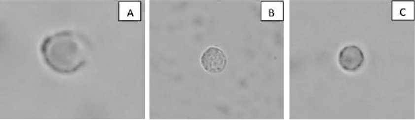
Figure 1. Morphological description of Blastocystis sp. on microscopic examination. A) Blastocystis sp. vacuolar form, B) Blastocystis sp. granular form, C) Blastocystis sp. cyst form (100x magnification).
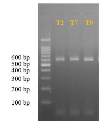
Figure 2. Visualization results of genus PCR products using electrophoresis on 1.5% agorose gel; M: TNAP markers and samples
Sampel Nt.2 - Alas Purwo-Indonesia
Sampel Nθ.19 - Alas PurwP-Indonesia
Sampel No.27 -Alas Purvrci-Indonesia
KX61S192.1_Singapore
MK416178.1-Turk⅛
MN33S083,1.lπ⅛
KY610153 I-Philipmes
OF270208J_lran
KY2 KMtM-I-MaIaysia
MGoiIMSJjranwi
OM478526.1_Uni_Emi rateJrab
OP942294. !.Egypt
Figure 3. Phylogenetic tree of Blastocystis sp. isolates in long-tailed macaques from Alas Purwo National Park, Banyuwangi, East Java
736
Discussion and feedback