Parasite Prevalence Oodinium sp. in Cantang Hybrid Grouper Cultivated in Recirculating Aquaculture System
on

Advances in Tropical Biodiversity and
Environmental Sciences
6(3): 79-84, October, 2022
e-ISSN:2622-0628
DOI: 10.24843/ATBES.2022.v06.i03.p03
Available online at: https://ojs.unud.ac.id/index.php/ATBES/article/view/88117
Parasite Prevalence Oodinium sp. in Cantang Hybrid Grouper Cultivated in Recirculating Aquaculture System
Kadek Leni Widiartini1, Kadek Lila Antara1, Ketut Mahardika2 and Gede Iwan Setiabudi1
-
1 Department of Aquaculture, Faculty of Mathematics and Sains, Ganesha of Education University Jl. Udayana No. 11, Banjar Tegal, Singaraja, Kabupaten Buleleng, Bali
-
2 Fisheries Research Center, Earth and Maritime Research Organization, National Research and Innovation Agency Jl. Singaraja-Gilimanuk, Banjar Dinas Gondol, Kabupaten Buleleng, Bali
*Corresponding author: leni@undiksha.ac.id
Abstract. The aims of this study were to determine clinical symptoms, mortality, prevalence, and histopathology of Cantang hybrid grouper fish infected with Oodinium sp. on the Cantang. The study was conducted by direct observation of clinical symptoms, calculating the number of fish mortality, and measuring the quality of water in the tank using the Cantang. While histopathology was conducted in the laboratory using 10 samples of sick fish with an average total length of 10.6 ± 0.69 cm and a weight of 18.6 ± 1.77 g. The results showed that the fish seen swimming weakly on the surface of the water near aeration or standing still at the bottom of the tub, decreased appetite, had pale or darker body color, and were thin. Observation of skin and gill mucus under a microscope showed the presence of ectoparasites Oodinium sp. in massive quantities in almost every gill sheet. The mortality of fish infected with Oodinium sp. in the Cantang of 26.84±3.9%, with a prevalence of 49.59%. Histopathologically the gill lamellae of fish infected with Oodinium sp. shows the occurrence of hyperplasia which causes the union of several gill lamellae.
Keywords: Cantang hybrid grouper fish, Recirculating Aquaculture System (RAS), Oodinium sp.
-
I. INTRODUCTION
Cantang hybrid grouper fish has the potential to be cultivated, because its growth relatively faster, easy to maintain, has a high tolerance for environmental changes and resistant to limited space such as being able to be cultivated in controlled containers or rearing tanks [17]. Grouper cultivation in a controlled system using RAS (Recirculating Aquaculture System) system. The system is an intensive fish farming system by using infrastructure that allows continuous use of water (water recirculation), using physical, chemical, and biological filters [4].
The application of the RAS in grouper fish aquaculture has begun to develop in Indonesia, although it is still on a research and trial scale in hatcheries. The Cantang for nursery fish such as high density white snapper (Lates calcarifer) has been successfully carried out at the Center for Marine Cultivation Research and Fisheries Extension (BBRBLPP), Buleleng Bali [10], although the population of Vibrio sp. bacteria and fish mortality is still being
monitored. Cultivation of Cantang hybrid grouper fish has also started in the Buleleng area, but there were still some obstacles, which hinder the development of aquaculture business. One of these inhibiting factors is infection with pathogenic microorganisms such as parasites.
Parasite that attack Cantang hybrid grouper fish is Oodinium sp. [19] The sympton can see from the fins with the presence of a powdery coating called velvet, the scales or fish skin peels off and the eyes would look like an opaque membrane. Furthermore, during cultivation the use of floating net cages reported to cause mass death due to damage to the skin and gills.
The cultivation of the Cantang in Indonesia found that there have not been reports of Oodinium sp. Therefore, this study aimed to obtain more information on clinical symptoms, mortality, prevalence, and histopathology of Cantang hybrid grouper fish infected By Oodinium sp. on the Cantang.
-
II. RESEARCH METHOD
Research Setting
This research had been conducted in one of the marine fish farming areas using the Cantang located in northern Bali, and at the Pathology Laboratory, Center for Marine Cultivation Research and Fisheries Extension (BBRBLPP), Gondol, Buleleng, Bali.
Fish Sample
The Cantang hybrid grouper sample obtained from the fish farm. Samples of fish tissue were collected from weak and dead fish. With an average total length of 10.6±0.69 cm and a weight of 18.6±1.77 g. The fish samples then fixed in 10% formalin solution.
Observation of Clinical Symptoms
Observation of clinical symptoms of Cantang hybrid grouper fish in this study conducted in fish rearing tanks using the Cantang. Observations made by direct observation of the swimming movements of the fish and the condition of the fish's body.
Ectoparasite Observation
Observations of ectoparasites were conducted on 10 moribund or recently dead fish. Fish samples placed in clean trays that had been sprayed with 70% alcohol. The surface of the skin or fish scales was scraped using a cover glass (size 24 x 24 mm) and placed on a glass object that had been dripped with sterile seawater. Fish gill lamellae were cut with scissors and placed on top of another glass object that had been dropped with sterile seawater. Then, the gill pieces covered with a cover glass. Observations of ectoparasites were conducted under a microscope with a magnification of 40-200 x.
Observation of Mortality and Prevalence of Oodinium sp.
Mortality observations were carried out by counting the number of fish that died every day while the fish were kept in the Cantang. Mortality observations carried out in 7 rearing tanks. Mortality is calculated using the formula:
number o f dea th
Mortality = -------f-----x 100%
,, number of spread
Prevalence observations were conducted by calculating the percentage of fish infected with the parasite divided by the number of fish samples examined. prevalence is calculated using the formula:
Prevalency
Number of fish infected by parasites
= -----H h -----X 100%
Number of fish checked
Histopathology
Histopathology had been conducted at the Pathology Laboratory of BBRBLPP Gondol. Gill samples of moribund or recently dead grouper fish hybrids taken from rearing tanks and fixed in 10% formalin for ± 2 weeks. Furthermore, the gill organs of the fish put into Bouin's solution for 24 hours to soften (decalcify) the bones in the gills. The histopathological process followed the procedure previously reported by [12] Histopathological preparations observed under a microscope with a magnification of 40-400x.
Water Quality
Observations of water quality were carried out during the incidence of infection due to the parasite Oodinium sp. Observations of water quality that are measured include: temperature, DO, pH, and salinity using a Photometer.
Data analysis
The data obtained during the study in the form of clinical symptoms, observations of ectoparasites, prevalence values, mortality values, and water quality data presented in the form of tables and graphs and then analyzed by qualitative and quantitative descriptive analysis by describing or describing the data that has been collected.
-
III. RESULTS AND DISCUSSION
Clinical Symptoms
Initial observations of the Cantang hybrid grouper fish showed of behavior changes. Sympton that occurred include a decrease in appetite where there was a decrease in response to feed when fish were fed commercial pellet feed. An increase in response to shock in which the fish were very sensitive to a movement caused by several other fish and the movement of people/workers, and swimming was not normal. It was because it sometimes swims weakly in the water column or stay at the bottom of the tub or swim weakly on the surface of the water near aeration (Figure 1A & B). These symptoms indicated the fish lack of oxygen (anorexia).
After 7 days after the appearance of clinical symptoms seen in fish behavior, the Cantang hybrid grouper fish also experienced physical changes. Physical changes were observed, including a change in the color of the fish (Figure 1C) where the color of the fish's body would slowly change as in fish fins which show a layer of flour called velvet, the scales or fish skin peels off and the eyes would look like an opaque membrane, The bodies of sick
fish mostly have body deformities, such as disproportionate body size between length and width, i.e. the length is far greater than the width so the fish looks very thin, slimy fish due to excessive mucus production as a defense for fish, and fish gills look red and red slimy (Figure 1D).
The clinical symptoms found in the Cantang hybrid grouper fish kept were the same as those found in koi fish infected By Oodinum sp. Rreported by [2] where on the results of examination of the body surface of the koi fish, it showed that some of the fish scales were starting to peel, swimming was not normal, and there was a lack of appetite [13] , also reported in his research on koi fish infected with Oodinium sp. has some red areas as a sign that the fish is infected with parasites and scales were peeling off. [19] reported clinical symptoms in fish infected with Oodinium sp. starting from the fish fins, advanced stages would look like wearing powder or sprinkled with flour, called velvet. In the next stage, the scales or skin of the fish would peel off, the eyes would see the opaque membrane and then attack all parts of the body. After the incident, the ectoparasite examination conducted with a microscope showed the presence of the parasite Oodinium sp. on skin mucus scrapings and fish gills.
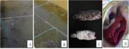
Figure 1. Clinical symptoms of Cantang hybrid grouper fish infected with Oodinium sp. A. fish appear darker in color and swim weakly near the surface of the water, B. fish that swim near aeration, C. Newly dead fish appear pale in color (bottom image) and/or blackish in color (top image) with excessive mucus production, D. Fish gills look reddish and slimy.
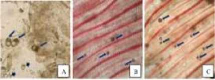
Figure 2. Cantang hybrid grouper fish infected with Oodinium sp. A. Scrapings of skin mucus containing Oodinium sp. adults (long arrow) and Oodinium sp. young (short arrows), B. Gill lamellae infested with a small number of parasites, C. Gill lamellae infested with a large number of parasites.
Fish Mortalities
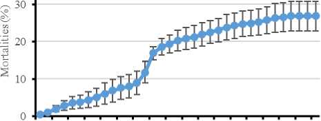
Death of Cantang hybrid grouper fish infected with Oodinium sp. approximately 2 months. During the ectoparasite infection, the fish treated using a solution of 1 ppm cupric sulfate (CuSO4) for daily treatment and 3 ppm for weekly treatment. During infection, every day the fish showing clinical symptoms, moribund and recently dead were immediately removed from the rearing tank to prevent the spread of infection from getting worse, and to reduce the mortality rate. In addition, to reduce mortality, maintenance water replacement is conducted. However, there was still a spike in deaths. The highest mortality occurred on day 15, this death was caused by fish that had been severely infected before. Mortality of Cantang hybrid grouper fish infected with Oodinium sp. in the Cantang, which is 26.84±3.9% (Figure 3). Fish mortality began to decrease after 32 days after infection which was marked by the restoration of fish appetite, fish swimming activities began to be active and fish body conditions began to fill with increasingly bright colors. According to [7] In Sunuk grouper fry in controlled tanks infected with Amyloodinium ocellatum, death occurred up to 28% within three days of initial symptoms. Mortality from parasites in the Cantang was low because the Cantang is more effective.
Ectoparasites Oodinium sp.
In skin mucus scrapings, it is seen the presence of Oodinium sp. with several different sizes. Oodinium sp. adults looked darker in color with a large size compared to Oodinium sp. young or at the dinosphore stage (Fig. 2A). Observation of gills showed the presence of a collection of Oodinium sp. which infects the secondary lamellae of the gills. Oodinium sp. It was round to slightly oval with a brownish color (Fig. 2B & C).
According to research conducted by [6] reported on the Sunuk grouper also found Oodinium sp. along the gill filaments and in the gill lamellae. The parasite looked round and oval with a red color resembling the color of the gills.
40
1 3 5 7 9 11 13 15 17 19 21 23 25 27 29 31 33 35
Emergence of infection (days)
Figure 3. Daily mortality of grouper that infected by Oodinium sp.
The prevalence of fish infected with Oodinium sp.
The prevalence of Cantang hybrid grouper fish cultured using the Cantang was 49.59%. The high prevalence of fish infected with Oodinium sp. can triggered by high stocking densities, causing the condition of fish to become susceptible to disease. This was also reported in research [10] who said that in tilapia cultivation in ponds, the prevalence caused by the parasite Oodinium sp. Amounted to 33.33%. Fish conditions can disturb due to high density, lack of nutrients and unsupportive water quality, which can cause fish to become weak and susceptible to disease.
Histopatology Symptons
Histopathologically on the gills infected by Oodinium sp. showed hyperplasia of the secondary lamellae. This was due to the presence of parasitic cysts between secondary lamella (Figure 4A-C). The gills infected By the Oodinium sp. resulting in fusion of gill lamellae due to secondary lamellae epithelial cell hyperplasia. As a result, the gill lamellae cannot function properly because the lacunae containing red blood cells are covered by secondary lamellae epithelial cells [18]. Furthermore, it reported that the gills with severe conditions would show the condition of the secondary gills with inter lamellae cells that would look like a bat. [19]
Sutarmat and Yudha [19] reported, Oodinium sp. caused by penetration of rhizoids into host epithelial cells, causing necrosis, hemorrhage and secondary infection by bacteria and fungi. Oodinium sp. would attach to the fish using a flagellum which would then form sucker rods (legs) that enter the mucous membrane of the fish gills. These sucker rods (legs) would damage the surrounding cells and suck up the nutrients in the fish flesh as food. According to [19] this can result in mass death due to damage to the gills.
Water Quality
The results of water quality measurements showed that water quality during rearing Cantang grouper fish cultured with the Cantang with the optimal conditions (Table 1). The water temperature during the study ranged from 29 – 31 C, indicating that the temperature was still in a suitable condition for rearing Cantang hybrid grouper fishes. According to SNI (2014:2) the optimal temperature for the growth of Cantang hybrid grouper fish is 28 – 32 C [8] reported on his research showing that the prevalence is related to water temperature, this can be seen with increasing temperature, the prevalence of parasites decreases. This was presumably because the optimal temperature range could support the fish's immune system so that it does not match the development of the parasites found, so that the parasites cannot adapt and reproduce in
increasing numbers. It was not suitable for the development of parasites that founded, so that these parasites were not able to adapt and reproduce. Therefore, the prevalence of parasites decreases as well.
The DO value or dissolved oxygen during the study was still in the safe range for rearing Cantang hybrid grouper fish, which was between 4.24 – 5.95 mg/l. According to SNI (2014:2), the optimum range of dissolved oxygen in Cantang hybrid grouper fish is >4. The degree of acidity or pH greatly affects the health of fish and the presence of parasites in the body of the fish. The results of the pH value during the study ranged from 7 to 7.89, the pH value during the study was normal and suitable for rearing Cantang hybrid grouper fish. According to SNI (2014:2), the optimum pH value for the growth of Cantang hybrid grouper fish is 28 – 33 ppt. The salinity value during the study ranged from 32 - 33 ppt, the salinity value during the study was normal and suitable for rearing Cantang hybrid grouper fish. According to SNI (2014:2), the optimum salinity value for the growth of Cantang hybrid grouper fish is 28 – 33 ppt.
Water quality was very influential on the survival of fish therefore it is necessary to maintain optimum water quality for fish so that fish would always be healthy and not stressed and not susceptible to disease or parasites. The average water quality during the study showed optimal conditions for rearing the Cantang hybrid grouper fish. In addition to water quality [10] stated that the condition of fish could be disturbed due to high density and lack of nutrients, causing the condition of the fish to become weak and susceptible to disease [4] High stocking density allowed the level of parasite attack on fish to be higher. This opinion was supported by the statement [4] that a high population would facilitate the transmission of parasites because the possibility of contact between sick fish and healthy fish would increase. Fish health management that was not carried out can trigger the emergence of parasites in the aquaculture environment. This affected the results of fish farming itself.
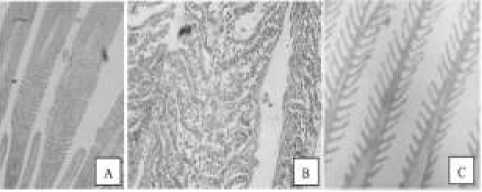
Figure 4. Histopathological description of the gills of the Cantang hybrid grouper fish infected with Oodinium sp. A.
The occurrence of hyperplasia in the secondary lamellae of the gills causes fusion (fused). Oodinium sp. seen between secondary lamellae, B. High enlargement of secondary gill
lamella showing hyperplasia, C. gill lamella from healthy fish.
TABLE I
WATER QUALITY PARAMETERS DURING REARING OF CANTANG HYBRID GROUPER IN THE RAS
|
Parameter of the Water Quality |
Water Quality Range during Research |
Optimal Range According to Library |
|
Temperature |
29 – 31 ْC |
28 – 32 ْC |
|
DO |
4,24 – 5,95 |
>4 |
|
Ph |
7 – 7,89 |
7,5 – 8,5 |
|
Salinity |
32 – 33 ppt |
28– 33 ppt |
-
IV. CONCLUSION
The clinical symptoms infection of Oodinium sp. in Cantang hybrid grouper fish cultured including behavioral and physical changes, the mortality rate caused by the parasite Oodinium sp. of 26.26% and prevalence of 48.84%, as well as histopathological of the gills of grouper fish showed severe damage to the gill organs.
ACKNOWLEDGEMENT
This contribution has been supported by Pathology Laboratorium of IMRAD. Gondol.
REFERENCES
-
[1] Abubakar, A. (2008). Prospek Penembangan Budidaya Ikan Kerapu Macan Sistem Keramba Jaring Apung (KJA) Di Gampong Mee Pangwa Kecamatan Trieggadeng Kabupaten Pidie Jaya. 41-53.
-
[2] Firdausi, A. P., Rahman, Mahadhika, R., & Sumadikarta, A. (2020). Protozoa Ektoparasitik Pada Ikan Koi Cyprinus Carpio Di Daerah Sukabumi. Jurnal Akuakultur Rawa Indonesia, 50-57.
-
[3] Handayani, Retna, Y.T. Adiputra, and Wardiyanto. “Identifikasi Dan Keragaman Parasit Pada Ikan Mas Koki (Carrasius Auratus) Dan Ikan Mas (Cyprinus Carpio) Yang Berasal Dari Lampung Dan Luar Lampung.” (1).
-
[4] Jacinda, A., Yustiati, A., & Andriani, Y. (2021).
Aplikasi Teknologi Resirculating Aquaculture System (RAS) Di Indonesia; A Review. Jurnal Perikanan Dan Kelautan, 11, 43-59.
-
[5] Mahardika, K., Mastuti, I., Roza, D., Syahidah, D., Astuti, W., Ismi, S., & Zafran. (2020). Pemantauan Insidensi Penyakit Pada Ikan Kerapu Dan Kakap Di Pembenihan Dan Keramba Jaring Apung Di Bali Utara. Jurnal Riset Akuakultur, 15 (2), 2020, 89-102.
-
[6] Mahardika, K., Mujimin, & Setyawati, K. M. (2017). Parasit Dinoflagelata (Amyloodinium Ocellatum) Pada Ikan Kerapu Sunu,
Plectropomus Leopardus. Seminar Nasional Kelautan XII, 71-77.
-
[7] Mahardika, K., Haryati, Muzaki, A., &
Miyazaki, T. (2008). Histopathological And Ultrastructural Features Of Enlarged Cells Of Humpback Grouper fish Cromileptes Altivelis Challeged With Megalocytivirus (Family Iridoviridae) After Vaccination. Disiases Of Aquatic Organisms, 79, 163-168.
-
[8] Mujimin, & Suratmi, S. (2018). Pengamatan Dan Penanganan Penyakit Parasit
Amyloodinium Ocellatum Pada Pendederan Benih Ikan Kerapu Sunu Di Bak Terkontrol. Buletin Teknik Litkayasa Akuakultur, 61-64.
-
[9] Manurung, U. N., & Saselah, J. T. (2017). Penyebaran Penyakit Parasit Pada Ikan Nila (Oreochromis Niloticus) Di Kabupaten Kepulauan Sangihe. Jurnal Ilmiah Tindalung, Volume 3, Nomor 1, Maret 2017, Hlm. 8–14.
-
[10] Manurung, U., & Gaghenggang, F. (2016). Identifikasi Dan Prevalensi Ektoparasit Pada Ikan Nila (Oreochromis Niloticus) Di Kolam Budidaya Kampung Hiung, Kecamatan Manganitu, Kabupaten Kepulauan Sangihe. Budidaya Perairan Mei 2016, 4, 26-30.
-
[11] Permana, Gusti Ngurah Et Al. 2019. “Aplikasi Sistem Resirkulasi Pada Pendederan Ikan Kakap Putih, Lates Calcarifer Kepadatan Tinggi.” Jurnal Riset Akuakultur 14(3): 173.
-
[12] Pratiwi, Harini Citra, And Abdul Manan. 2015. “Teknik Dasar Histologi Pada Ikan Gurami (Osphronemus Gouramy) The Basic Histology Technique Of Gouramy Fish (Osphronemus Gourami).” Jurnal Ilmiah Perikanan Dan Kelautan 7: 153–58.
-
[13] Priawan, Indra, Endang Sulistyarini Gultom, and Ahmad Shafwan S Pulungan. 2017. “Identifikasi Ektoparasit Pada Ikan Koi ( Cyprinus caprio ).” 3(1): 21–24.
-
[14] Sari, D. R., Sarjito, & Desrina. (2021). Efektivitas Perendaman Perasan Biji Pepaya (Carica Papaya L.) Untuk Mengendalikan Infestasi Argulus Sp. Pada Ikan Mas (Cyprinus Carpio L.). Jurnal Sains Akuakultur Tropis, 8087.
-
[15] Saselah, Jetti T, And Usy N Manurung. 2017. “Penyebaran Penyakit Parasit Pada Ikan Nila (Oreochromis Niloticus) Di Kabupaten Kepulauan Sangihe.” Jurnal Ilmiah Tindalung 3(1): 8–14.
-
[16] Septinawati, A., & Tjahjaningsih, W. (2010). Manajemen Pembesaran Kerapu Tikus
(Cromileptes Altivelis) Di Balai Besar Pengembangan Budidaya Air Payau (BBPBAP) Jepara Jawa Tengah. Jurnal Ilmiah Perikanan Dan Kelautan, 2, 67-75.
-
[17] Sudaryatma, P. E., & Eriawati, N. (2012). Histopatologis Insang Ikan Hias Air Laut Yang Terinfeksi Dactylogyrus Sp. Jurnal Sain
Veteriner, 68-75.
-
[18] Suratmi, Sri, And Bahan Alat. 2018. Pada Pendederan Benih Ikan Kerapu Sunu Di Bak Terkontrol. 16(1): 61–64.
-
[19] Sutarmat, T., & Yudha, H. T. (2013). Analisis Keragaan Pertumbuhan Benih Kerapu Hibrida Hasil Hibridisasi Kerapu Macan (Epinephelus Fuscoguttatus) Dengan Kerapu Kertang
(Epinephelus Lanceolatus) Dan Kerapu Batik (Epinephelus Microdon). Jurnal Riset Akuakultur, 8, 363-371.
-
[20] Utami, I. S., Ciptojoyo, A. A., & Wiadnyana, N. N. (2017). Histopatologi Insang Ikan Patin Siam (Pagasius Hypophthalamus) Yang Terinfestasi Trematoda Monogenea. Media Akuakultur , 3543.
-
[21] Wirawan, I Kadek Adi, Sang Ayu Made Putri Suryani, And I Wayan Arya. 2018. “Diagnosa, Analisis Dan Identifikasi Parasit Yang Menyerang Ikan Nila (Oreochromis Niloticus) Pada Kawasan Budidaya Ikan Di Subak ‘Baru’ Tabanan.” Gema Argo 23(1): 6.
Discussion and feedback