Lung Histopathology of Laying Hens Infected by Colibacillosis in the Animal Cages Experiments of the Disease Investigation Center 6, Denpasar, Bali
on
Advances in Tropical Biodiversity and Environmental Sciences 3(2): 25-28, September 2019 ISSN : 2622-0628
DOI: 10.24843/atbes.v03.i02.p02 Available online at: https://ojs.unud.ac.id/index.php/ATBES/article/view/53813
25
Lung Histopathology of Laying Hens Infected by Colibacillosis in Animal Cages Experiments in the Disease Investigation Center 6, Denpasar, Bali
Kadek Ayu Trisna Yanti, Iriani Setyawati*, and Ni Putu Adriani Astiti
Biology Study Program, Faculty of Mathematics and Natural Sciences, Udayana University, Kampus Bukit Jimbaran, Badung, Bali *Corresponding author: iriani_setyawati@unud.ac.id
Abstract. This study aimed to determine the lungs histopathology of laying hens (Gallus gallus domesticus) at the Animal Cage Experiments in the Disease Investigation Center 6, Directorate General of Live Stock (DIC-6 DGLS), Denpasar, Bali, which died from colibacillosis infection. Sample of lungs were cut transversely then put into 10% of Neutral Buffer Formalin, then processed histologically by paraffin method and stained with Hematoxylin-Eosin. Observation under microscope (magnification 100x and 400x) was done for histopathological examination. Laying hens died from colibacillosis infection showed that their lungs were infected by colibacillosis, and there were found 62.50% of necrosis, 75% of inflammatory cells infiltration and 80% of hemorrhage in the lungs.
Keywords: colibacillosis; hemorrhage; laying hens, lung histology; necrosis.
-
I. INTRODUCTION1
Laying hens, one of favorite commodity, can produce a rapid capital turnover, because the production results (eggs) are favored by many Indonesian people. Laying hens produce eggs almost every day and almost 250-280 eggs per year. The eggs are the perfect food ingredients for toddlers to adults, it contains important nutrient such as protein, vitamins, fat, and minerals in sufficient quantities [1]. The maintenance period of laying hens generally lasts for 2 years and the production period starts at the age of 20 weeks or around 5 months [2].
A long maintenance period with polluted cages allows poultry to be attacked by many diseases. Colibacillosis or colisepticemia is an infectious disease in poultry caused by Escherichia coli, a pathogenic bacteria as primary or secondary agents. This E. coli infection can occur in broilers and laying hens of all age groups, as well as other poultry such as turkeys and ducks [3].
The clinical signs of colibacillosis are nonspecific and are influenced by the age of chickens, duration of infection, organ attacks, and the presence of other diseases. Broiler chickens aged 4-8 weeks and laying hens around 20 weeks can get an acute colibacillosis which cause death, generally begins with loss of appetite, inactive condition (lazy to move) and sleepy [4]. Colibacillosis transmission usually occurs orally through
food, drinking water, dust or dirt (feces) contaminated by E. coli. Dust in a chicken coop can contain 105-106 E. coli/gram and this bacteria can last a long time, especially in dry conditions. If dust is inhaled by chickens, it can infect the respiratory tract [5].
Colibacillosis can be explained in terms of organ abnormalities including septicemia, enteritis, granulomas, and airsacculitis [6]. Colibacillosis has an important economic significance for the poultry industry, because it can cause growth disturbances, increase the number of rejected chickens, decrease production and also the quality of the chicks, carcasses and eggs. In addition, the presence of E.coli infection can be a supporting factor for the emergence of complex respiratory, digestive or reproductive diseases that are difficult to overcome [5].
This study aimed to determine the lungs histopathology of laying hens (Gallus gallus domesticus) at the Animal Cage Experiments in the Disease Investigation Center 6, Directorate General of Live Stock (DIC-6 DGLS), Denpasar, Bali, which died from colibacillosis infection.
-
II. RESEARCH METHODS
Five dead laying hens, aged about 20 weeks, from the Animal Cages Experiments in the DIC-6 DGLS Denpasar were first put into the bacteriology laboratories of the DIC-6 DGLS Denpasar, Bali. The examination under a veterinary supervision at the bacteriology laboratory found Escherichia coli bacteria in animal organs that could be
diagnosed as a cause of colibacillosis.
Dead animals then were taken to the pathology laboratory to be processed into histological preparations. The animals were moistened under running water then were dissected to collect the lungs. The lungs were cut in an upright position transversely with a size of 2x1x1.5 cm, then were put into a jar contained with 10% of Neutral Buffer Formalin. The lungs then were processed with a tissue processor (Tissue Tek) to produce paraffin blocks.
The paraffin blocks were sectioned with a rotary microtome at a thickness of 5 μm. After an affixing process, the sliced tape of organs were stained with Hematoxylin-Eosin staining and then covered (in a mounting process) with cover glass and adhesive (permount). The preparation methods followed the standard operational procedure of DIC pathology laboratory. The histology slides were then observed under a light microscope and documented.
-
III. RESULTS AND DISCUSSION
Results
Based on the observations, the results obtained in the form of anatomical damage of the lungs that show hemorrhage (tissue bleeding) (Figure 1) and the average number of histopathological damage namely necrosis, inflammatory cells infiltration and hemorrhage (Table 1).
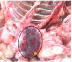
Fig 1. Lungs anatomy of Gallus gallus domesticus; a. hemorrhage
TABLE 1.
PERCENTAGE OF HISTOPATHOLOGICAL DAMAGE OF GALLUS LUNG DUE TO COLIBACILLOSIS INFECTION
|
Histopathological Damage |
Percentage of Damag |
e (%) D |
Average (%) | ||
|
A |
B |
C | |||
|
Necrosis |
60 |
70 |
60 |
60 |
62.50 |
|
Inflammatory cell infiltration |
80 |
90 |
55 |
75 |
75 |
|
Hemorrhage |
90 |
80 |
60 |
90 |
80 |
Note: A, B, C, and D are replications
Table 1 showed that the average percentage of histopathological damages of chicken lungs infected with colibacillosis were the occurrence of 62.50% necrosis (cell
death), 75% of inflammatory cells infiltration, and 80% of hemorrhage in the lung. Lungs histopathology of laying hens (Gallus gallus domesticus) showed there were necrotic cells, inflammatory cells (leucocytes) infiltration, and hemorrhage (Figure 2).
Discussion
The chicken lungs is usually orange-red and sponge-like macroscopically, and can be filled with air very well. If the lungs undergo pathology due to bacterial infection, it will cause inflammation of the lungs, the color will be red brown like a flesh and heavy [7]. Figure 1 showed that there was hemorrhage on the surface of the lungs which was reddish brown. According to Suyono [8], hemorrhage in the lungs occurs due to an infection of microorganisms found in the mouth, nose and airways. The main signs of acute inflammation due to colibacillosis including swelling or edema, hemorrhage, heat and malfunctioning of organs that are already severe [9].
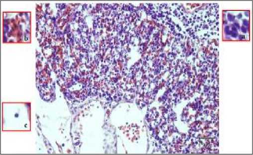
Fig 2. Histology of Laying Hens (Gallus gallus domesticus); a. Inflammatory Cell Infiltration, B. Hemorrhage, C. Picnotic Nucleus (Necrosis), Magnification 400x.
On microscopic observation of respiratory system (lungs) infected with colibacillosis, namely congestion, hemorrhage, neutrophil cell infiltration from the lumen of the bronchioles to the alveolar lumen and many necrosis occur in the septa alveoli [10]. In this study, histopathological identification on chicken lung due to colibacillosis infection found 62.50% of necrosis (cell death), 75% of inflammatory cells infiltration, and 80% of hemorrhage (Table 1, Figure 2).
The results in Table 1 showed that the necrotic damage of 62.50% means that the damage was already severe. Necrosis is one of the basic patterns of cell death. Necrosis usually occurs after the cells were exposured to toxins and were characterized by cell swelling, protein denaturation and organelle damages. This can cause severe tissue dysfunction [11]. According to Kardena et al. [12], necrosis is a cell death that can continue to tissue death. If it occurs in an organ, it can cause discoloration
of the organ. The aspect of color change depends on the type of necrosis that accompanies it. Usually, organs whose tissues or their constituent cells die, will be paler or darker in color than the normal color of the organ.
Table 1 and Figure 2 showed the presence of 75% inflammatory cell infiltration which means that the damage has been severe. Infiltration of inflammatory cells is a condition where the body tries to repair itself by involving the body's defense system when exposed to foreign antigens or injury. Inflammation is generally grouped into two, namely those caused by microorganisms (viruses, bacteria, parasites and other microorganisms) and nonmicroorganisms (chemicals, extreme temperatures, trauma and others) [14].
Inflammation that occurs in cases of colibacillosis is characterized by the presence of neutrophil inflammatory cells on microscopic examination. The severity of inflammatory cells infiltration is also influenced by the length of time the inflammation occurs, while subacute inflammation will decrease in severity. This could happen because cytokines stimulate an increase in neutrophil segments into the blood circulation. The severity of inflammatory cells infiltration is also influenced by the number of foreign agents, for example a bacterium that infects a tissue in an individual. The more foreign agents that enter the body, the more the response of inflammatory cells will be seen in the inflammatory process [15].
Table 1 and Figure 2 also showed that hemorrhage (tissue bleeding) in chicken lung organs by 80%, while damage of more than 75% indicates that the damage in the chicken lung organs has been very severe. Pathological hemorrhage is characterized by the presence of red blood cells outside the blood vessels or in tissues. On microscopic examination, hemorrhage is characterized by the presence of blood outside the blood vessels, indicated by the presence of red cells (erythrocytes) especially in Hematoxylin and Eosin staining [14].
Hemorrhage is caused by the effects of E. coli enterotoxin which causes an increase vascular permeability so that red blood cells come out of the blood vessels. The cause of the lesions in pulmonary histology is due to bacterial enterotoxin resulting in degeneration of the heart muscle until there is disruption of blood circulation from the heart. In this situation the blood can be blocked in the lungs. If this situation lasts a long time, it will result in hemorrhage [7].
Transmission of colibacillosis usually occurs orally through food, drinking water, and dust or dirt contaminated by E. coli. Dust in chicken coops can contain 105-106 E. coli/ gram and this bacterium can last a
long time, especially in dry conditions. If the dust is inhaled by chickens, it can infect the respiratory tract [5].
When chickens suffer from respiratory diseases caused by dust containing E.coli and inhaled through the respiratory tract, E.coli will enter the respiratory tract after inhalation. This bacteria will be attached to the surface of the respiratory tract epithelial. The specific attachment of this bacteria is due to the presence of villi. The bacteria then will enter the bloodstream and make multiplication in the chicken's body and eventually cause damage.
Significant damage will occur along the respiratory tract (trachea, lungs, and air sacs), and also pericardium and peritoneum. E. coli infection can also spread to other organs such as kidneys, liver, heart, and air sacs in the abdomen, make the body to be covered with fibrin. Chickens that experience are growth stunted and difficult to be treated, so the mortality rate becomes 8-10% [13].
-
IV. CONCLUSION
Chicken lung organ infected with colibacillosis showed necrosis in the form of 62.50%, infiltration of inflammatory cells by 75% and hemorrhage by 80%.
REFERENCES
-
[1] Indrawan IG., I M. Sakada, dan I K. Suada. 2012. Kualitas Telur dan Pengetahuan Masyarakat tentang Penanganan Telur di Tingkat Rumah Tangga. Denpasar.
-
[2] Zulfikar. 2013. Manajemen Agribisnis dan Pengolahan Hasil Peternakan. Badan Penyuluh Pertanian (BPP) Kabupaten Bireuen.
-
[3] Charlton, B.R., A.J. Bermudez, D.A. Halvorson., J.S.Jeffrey., L.J. Newton, J.E. Sander and P.S.Wakernell. 2000. Avian Diseases Manual. Fifth Edition. American Association of Avian Pathologist. USA: Poultry Pathology Laboratory University of Pennsylvania. New Bolton Center.
-
[4] Gomis, S.M., A.I. Gomis., N.U. Horadagoda, T.G. Wijewardene, B.J. Allan and A.A. Potter. 2000. Studies on Cellulites and Other Disease Syndromescaused by Escherichia coli in Broilers in Sri Lanka. Trop. Anim. Health Prod. 32(6): 341-351.
-
[5] Tabbu, C.R. 2000. Penyakit Ayam dan Penanggulangannya. Vol. I. Yogyakarta: Penerbit Kanisius.
-
[6] Akoso, B.T. 1993. Manual Kesehatan Unggas. Edisi Pertama.Yogyakarta: Kanisius.
-
[7] Meha, H.K.M., I.K.Berata., dan I.M.Kardana. 2016. Derajat Keparahan Patalogi Usus dan Paru Babi
Penderita Kolibasilosis. Indonesia Medicus Veterinus. 5(1): 13-22.
-
[8] Suyono, S. 2001. Buku Ajar Ilmu Penyakit Dalam Jilid II Edisi 3. Jakarta: Balai Penerbit FKUI
-
[9] Price, S and L. Wilson. 1995. Fisiologi Proses-Proses Penyakit Edisi 4. Alih Bahasa P.Anugrah. Jakarta: EGC.
-
[10] Dharma, D.M.N dan A.A.G. Putra. 1997. Penyidikan Penyakit Hewan. Denpasar: CV Bali Media.
-
[11] Kumar, V., R.S. Cotran, dan S.L. Robbins. 2007. Buku Ajar Patologi 7nd Ed, Vol. 2. Jakarta: Penerbit Buku Kedokteran EGC.
-
[12] Farooque, A.M.D., A. Mazunder, S. Shambhawee, R. Mazumder. 2012. Review on Plumeria acuminata.
Int. J. Res. Phar. Chem. 2:2
-
[13] Kardena, I.M., I.B.O.Winaya., I.K. Berata. 2011. Gambaran Patologi Paru-paru pada Anjing Lokal Bali yang Terinfeksi Penyakit Distemper. Buletin Veteriner Udayana. 3(1): 17-24.
-
[14] Jahja, J., C.L. Lestariningsih, N. Fitria, T. Muwrijayati, dan T. Suryani. 2006. Penyakit Penyakit Penting pada Ayam Edisi 5. Bandung: Medion.
-
[15] Berata, I.K, I.B.O. Winaya, A.A.A Mirah Adi, dan I.B.W. Adnyana. 2011. Patologi Veteriner Umum. Denpasar: Swasta Nulus.
-
[16] Rahmawandani, F.I. 2013. Studi Patologi Kasus Kolibasilosis Pada Babi Landrace Berdasarkan Umur. Denpasar. FKH Universitas Udayana.
Discussion and feedback