Labia and Lingua Histopathology of Bali Cow (Bos sondaicus) on Hepatogenous Photosensitization Symptoms in Pakutatan Village, Jembrana, Bali
on
Advances in Tropical Biodiversity and Environmental Sciences 2(2): 31-36, September 2018
ISSN : 2622-0628
31
Labia and Lingua Histopathology of Bali Cow (Bos sondaicus) on Hepatogenous Photosensitization Symptoms in Pakutatan Village, Jembrana, Bali
Kadek Mardika, Iriani Setyawati* and Dwi Ariani Yulihastuti
Biology Study Program, Faculty of Mathematics and Natural Sciences, Udayana University Kampus Bukit Jimbaran, Badung, Bali
*Corresponding author: iriani_setyawati@unud.ac.id
Abstract. Hepatogenous photosensitization is one of the ruminant diseases with symptoms of dermatitis or eczema of the skin accompanied by liver damage. The disease is caused by the compounds of toxic lantadene A and lantadene B which are secondary metabolites of Lantana camara plant. This research was carried out on January 2017. The material used in this study was preserved organs of the labia and lingua of dead three year old cow (Bos sondaicus). Samples were taken from Pakutatan Village, Jembrana, Bali. Identification of organ samples, histological preparation and histopathological examination were conducted at the Disease Investigation Center (DIC) 6, Denpasar, Bali. The preparation of labia and lingua samples used the paraffin method with Hematoxylin and Eosin staining. The tissue structural damages found were necrosis, inflammatory cell infiltration, congestion and tissue bleeding. The data obtained were analyzed statistically by One Way Anova test with a confidence level of 95%. Based on the results, dead Bali cow which consumed a large numbers of Lantana camara plants showed that the highest number of cell damage was at the picnotic stage of cell necrosis (cell death) in the labia organ significantly (P<0.005), while the highest bacterial invasion was found in the labia organ with an average percentage of 12.40%.
Keywords: photosentization, Balinese cow, Lantana camara, lantadene, histopathology
-
I. INTRODUCTION1
Continuous, high-quality, inexpensive, and practical feed supply is sought by farmers to get better results. Problems encountered in providing food in general are low quality feed, limited sources of feed, and the distance between feed sources and livestock [1]. In addition, the lack of knowledge of breeders in the selection of feed causes livestock to be poisoned by toxic compounds produced by several plants. Toxic compound poisoning can cause various kinds of diseases, such as hepatogenous photosensitization, known as Bali Ziekte. Hepatogenous photosensitization is a disease that commonly attacks ruminants including cows, sheep, goats and horses. Early symptoms of the disease are in the form of dermatitis or eczema of the skin and accompanied by damage to liver hepatocyte cells [2]. Hepatogenous photosensitization was first reported in New Zealand in 1833 in the form of skin eczema found in sheep. Whereas in Indonesia the symptoms of hepatogenous photosensitization have been found since 1925 in Bali cattle with the same symptoms, namely eczema on the skin (skin eczema) [3].
Hepatogenous photosensitization found in 3 years old Bali cow (Bos sondaicus) that died on 3 January 2017 in Pakutatan Village, Pakutatan District, Jembrana, Bali. Symptoms appeared were dermatitis or lesions on the nose, mouth, head, ears, back and legs. The internal organs, the lymph and the liver, were swollen (edema), the gallbladder enlarged, and the renal cortex was dark in color. It happened because the cow accidentally consumed Lantana camara plants in large quantities. This plants contained lantadene A and lantadene B compounds [4].
Lantana camara (Verbenaceae family) is a shrub that has a distinctive odor and has a trichular cytoma on the entire surface of its organs, and is widely grown in tropical and subtropical regions [5]. This plant is classified as 10 dangerous weeds in the world [6]. In Pakutatan Village, Jembrana, Bali, the plants thrive and are usually used by local people as plant biopesticides.
Labia and lingua organs of Bali cow who died due to poisoning of lantadene A and B compounds, were examined at the Disease Investigation Center (DIC) 6 Denpasar. The examination was needed to diagnose or ascertained whether the cause of death of Bali cow was purely caused by the toxic compound of Lantana camara.
-
II. RESEARCH METHODS
The research was conducted on January 2017. Trimming and preservation of the samples, sectioning, staining, and mounting preparations, followed by histopathological examination were carried out in the Pathology Laboratory of the DIC 6 Denpasar.
Histological Preparation
The histological preparation method followed the standard procedure of the Pathology Laboratory in DCG 6 Denpasar. Fresh labia and lingua of the Bali cow were fixed in 10% of formalin buffer solution. Embedding cassettes contained the tissues were put into a tissue processor (Tissue Tech) that worked automatically for 18 hours, the temperature was set at 650C and the cryo temperature was -50C. After that, the labia and lingua were taken from the embedding cassettes, then transferred to the paraffin block and liquid paraffin was added. The paraffin block was placed in cryo for several minutes so that the paraffin block becomes solid.
The sectioning process was carried out with the rotary microtome and the slides thickness was set at 5 µm. The sliced tape obtained from the sectioning process was placed into a floating bath (with a temperature of 380C) for a few minutes for stretching. After being incubated in an incubator (460C) for 30 minutes, then the affixing process can be applied to the paraffin tape.
The tissue slides were dipped in a xylol solution (deparaffinization), a series of alcohol concentration (dehidration), and washed with distilled water. The slides were stained with Hematoxylin for 15 minutes, dipped in
acid alcohol, washed in distilled water and then put into Eosin for 3 minutes. After staining, the slides were dipped in alcohol (96% and 100%) and xylol. After drying for several minutes, the mounting process was done using permount solution (as adhesive), so that the glass cover with the stained tissues can adhere well.
Histopathological Observation
The histopathology of labia and lingua of Bali cow were observed using a light microscope (Olympus BX-51) with a magnification of 400. Cell damages were found by looking at the structure of cell nucleus and vacuole. The number of damage cells were counted with the Image Raster software (Optilab by Micronos) and calculated in each field of view under microscope observation.
The damages of the histological tissue found in labia and lingua in this observation were cell necrosis (nucleus pycnotic, karyorrhexis, karyolysis, and cell lysis), congestion (blockage of blood vessels), tissue bleeding, and bacterial invasion, found in the layer of sub muscularis and sub mucosa, and also hair follicles. The data were analysed statistically using One Way Anova test with a confidence level of 95%.
-
III. RESULTS AND DISCUSSION
Results
Labia Histopathology of Bali Cow
Identification of the labia histopathology was done by comparing the normal and the exposured condition to the toxic compounds of Lantana camara based on the average number of cell damages including cell necrosis, congestion, bleeding and bacterial invasion (Table 1).
TABLE 1.
HISTOPATHOLOGY OF CELL NECROSIS IN THE LABIA OF BALI COW DUE TO POISONING OF Lantana camara
|
Group |
Cell Average of Necrosis Phase | |||
|
Pycnotic |
Karyorrhexis |
Karyolysis |
Lysis | |
|
Unexposed |
0 ±0 a |
0 ±0 a |
0 ±0 a |
0 ±0 a |
|
Exposed |
98.2 ±55.03 b |
14.0 ±10.29 b |
10.4 ±2.79 b |
17.2 ±6.53 b |
Different superscript letters in the same column show significant differences (P<0.005).
TABLE 2.
HISTOPATHOLOGY OF CONGESTION AND BACTERIAL INVASION IN THE LABIA OF BALI COW DUE TO POISONING OF Lantana camara
|
Group |
Average Percentage (%) | |
|
Congestion |
Bacterial Invasion | |
|
Unexposed |
6.9 ± 10.02 a |
0 ±0 a |
|
Exposed |
25.54 ± 20.36 a |
12.40 ± 14.02 a |
Different superscript letters in the same column show significant differences (P<0.005).
Table 1. showed that in the unexposed group there were no cells that experienced histopathological damage leading to necrosis (cell death), whereas the average number of cells of the exposed group that suffered cell damage in the form of picnotic nuclei (shrinking and compacting cell nuclei) was 98.2 ± 55.03 cells, karyorrhexis nucleus (breakage of the nucleus and chromatin damage) was 14.0 ± 10.29 cells, karyolysis nucleus (dissolution of chromatin in the nucleus) was 10.4 ± 2.79 cells, and lysis cells (ruptured cell membranes and exit cell organelles) was 17.2 ± 6.53 cells. Statistical test results with One Way Anova showed a significant difference (P<0.005) between
the unexposed and exposed group.
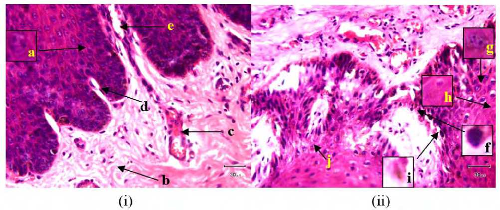
Table 2. showed that the average percentage of congestion (abnormal accumulation in blood vessels or body channels) in the unexposed group was 6.9 ± 10.02 while in the exposed group was 25.54 ± 20.36%. Bacterial invasion in the unexposed was not found while in the exposed found bacterial invasion with an average percentage of 12.40 ± 14.02. Statistical test results with One Way Anova showed non significant differences (P>0.05). Histopathological features of the labia cells between the unexposed and exposed groups were different as shown in Figure 1.

Fig.1. Histology of Balinese cow labia organ
(i) unexposed (normal), (ii) exposed to the toxic compound of Lantana camara; a. normal epithelial cell nucleus, b. lamina propria, c. blood vessels congestion, d. lamina propria of mucosal layer, e. Blood vessels, f. pycnotic nucleus, g. karyorrhexis nucleus, h. karyolysis nucleus, i. pre-lytic cell, j. bacterial invasion (Magnification 400x).
Lingua Histopathology of Bali Cow
Identification of the labia histopathology of the Bali cow was done by comparing the normal and the exposured condition to the toxic compounds of Lantana camara plant based on the average number of cell damages including cell necrosis, congestion, bleeding and bacterial invasion (Table 3 and Table 4).
Table 3. showed that the average number of cells of the exposed group that suffered damage to the picnotic nucleus was 102.60 ± 44.83 cells, the karyorrhexis phase was 18.40 ± 16.53 cells, the karyolysis phase was 19.20 ± 19.40 cells, and the cell lysis was 5.80 ± 8.55 cells. The results of statistical tests with One Way Anova only on the parameters of pycnotic (P = 0.001) and karyorrhexis (P = 0.038) showed a significant difference (P<0.005) between
the unexposed group and the exposed group. Other parameters showed non significant differences between unexposed and exposed group.
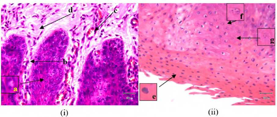
Table 4. showed that the average percentage of congestion of blood vessels in the unexposed was 6.9 ± 10.02 while in the exposed it was 25.54 ± 20.36. The average percentage of bacterial invasion and bleeding were not found in the unexposed whereas in the exposed group the average percentage of bleeding and bacterial invasion was 24.40 ± 32.90 and 8.40 ± 11.69, respectively. Statistical analysis with One Way Anova test showed non significant differences (P>0.05) between the unexposed group and the exposed group. The description of lingua cell histopathology between the unexposed and exposed groups was different as shown in Figure 2.
TABLE 3.
HISTOPATHOLOGY OF CELL NECROSIS IN THE LINGUA OF BALI COW DUE TO POISONING OF Lantana camara
|
Group |
Cell Average of Necrosis Phase | |||
|
Pycnotic |
Karyorrhexis |
Karyolysis |
Lysis | |
|
Unexposed |
5.0 ±7.54 a |
0 ±0 a |
0 ±0 a |
0 ±0 a |
|
Exposed |
102.60 ±44.83 b |
18.40 ±16.53 a |
19.20 ±19.40 a |
5.80 ±8.55 a |
Different superscript letters in the same column show significant differences (P<0.005).
TABLE 4.
HISTOPATHOLOGY OF CONGESTION, BLEEDING, AND BACTERIAL INVASION IN THE LINGUA OF BALI COW DUE TO POISONING OF Lantana camara
|
Group |
Average Percentage (%) | ||
|
Congestion |
Bleeding |
Bacterial Invasion | |
|
Unexposed |
6.9 ± 10.02 a |
0 ±0 a |
0 ±0 a |
|
Exposed |
25.54 ± 20.36 a |
24.40 ± 32.90 a |
8.40 ± 11.69 a |
Different superscript letters in the same column show significant differences (P<0.005).

Fig.2. Histology of Balinese cow lingua organ
(i) unexposed (normal), (ii) exposed to the toxic compound of Lantana camara; a. normal epithelial cell nucleus, b. lamina propria of mucosal layer, c. blood vessels congestion, d. lamina propria, e. piknotic nucleus, f. karyorrhexis nucleus, g. karyolysis nucleus (Magnification 400x).
Discussion
Histopathological identification of the labia and lingua of the dead Bali cow due to exposure to toxic compounds of Lantana camara plant showed more cell nucleus that experienced necrotic (cell death) of the pycnotic phase and followed by karyorrhexis, karyolysis, and lysis phases of the cells.
Necrosis is one of the basic patterns of cell death. Necrosis occurs after blood supply is lost or after exposure to toxins and is characterized by cell swelling, protein denaturation and organelle damage. This can cause severe tissue dysfunction [7].
The average number of cells that suffered necrotic damage was found more in the lingua organ (Table 3) compared to the labia (Table 1) of Bali cow. Lantana camara plant contains toxic compounds namely flavonoids, lantadene A and B, diterpenoids and triterpenoids. According to Toit [8], lingua functions as a digestive organ by facilitating the movement of food during mastication and helps swallowing food.
Lingua is the first organ that is in direct contact with the lantadene A and B compounds, assisted by cellular trichomes in the mechanical process of digestion. In addition, flavonoid compounds help in increasing the
permeability of cell walls to facilitate the lantadene A and B compounds to enter into the cell [9].
Beside lantadene, phylloerythrin is a metabolic product that is also toxic to the body of ruminant animals so it needs to be excreted through the gallbladder [10]. Lantadene compounds can cause thickening of the bile canaliculi due to epithelial cells of the gallbladder duct undergoing proliferation so that phylloerythrin which should be excreted into the gallbladder by the liver will be distributed into the peripheral blood vessels [11].
The dead Bali cow labia organ showed more necrotic cell damages in the lysis phase compared to the lingua organ which was an average of 17.2 ± 6.53 cells while in lingua as many as 5.80 ± 8.55 cells. The number of cells undergoing lysis in the Bali cow labia was assumed due to the excessive accumulation of toxic metabolites, phylloerythrin and lantadene, inside the cells and tissues.
Cells that store phylloerythrin and lantadene compounds will be active when exposed to direct sunlight, cause the skin that is not covered by hair looks like cracked. Cow skin which undergoing hepatogenous photosensitization showed epithelial cells become lysis so that they were detached from the connective tissue. This was supported by the statistical test results that showed a real difference (P=0.00) to the unexposed, that the toxic compounds lantadene A and B affected the cell damage of the Balinese cow labia due to the large amount of animal feed in the form of Lantana camara plants in Bali cattle (Simantri Program). Sani [12] reported that the toxic dose of lantadene A in sheep was 60 mg/ kg orally and 4 mg/ kg intravenously. The leaves are the main source of the lantadene A toxin in the Lantana camara plant, while the flowers, fruits and roots contain very low toxins and do not cause liver and bilirubin abnormalities in guinea pigs.
Lantana camara also contains a compound of diterpenoid (C20), a toxic compound, that has 20 number of carbon atoms. These compounds have the potential to irritate and inhibit cell growth, including skin cells (stratified squamous epithelium) in the labia organ so that it becomes disrupted in division to regenerate dead cells.
Triterpenoid compound is a compound that has antibacterial activation so that bacterial invasion was lower in the lingua compared to the labia of bali cow [13]. In this research, bacterial invasion had an average percentage of 8.40 ± 11.69 in the lingua of dead Bali cow by consuming Lantana camara plant. The small amount of bacterial invasion in the lingua was also caused by the saliva. Saliva plays role as an antibacterial by lysing or destroying certain bacteria in the lingua, then rinsing the material that the bacteria might use as food source and neutralizing the acidic atmosphere (pH<7) which was produced by bacteria in the mouth [14].
In a normal condition in the labia, there are various kinds of bacteria that are part of the normal flora called apatogens, which do not cause any negative impact. Hepatogenous photosensitization can cause a decrease endurance of the labia or body of Bali cow, so apatogenic bacteria will become pathogenic and cause disruption or cause various diseases or infections [15]. Statistical test results showed that bacterial invasion in the labia of exposed group had an average percentage of 12.40 ± 14.02 but not significantly different from the unexposed (P=0.084).
There were found congestion (Figures 1 and 2) and bleeding (Figures 2) in the labia and lingua of the exposed group. The feeding of Lantana camara leaves in Bali cow caused histological damage in the form of blood vessels congestion, but there was no significant difference between exposed (P labia = 0.104; P lingua = 0.139) and unexposed group. Congestion is an increase in blood cells in tissues and parts of the body undergoing a pathological process [16]. Toxic substances that enter the body will disrupt the circulation system so that oxygen and food nutrient can not be processed in the body [17].
-
IV. CONCLUSION
Dead Bali cow which consumed a large numbers of Lantana camara plants showed a significant number of pycnotic nucleus (cell necrosis) in the lingua, while the highest bacterial invasion was found in the labia with an average percentage of 12.40%.
REFERENCES
-
[1] Trisyulianti, E., J. Jachja, dan Jayusmar. 2001. Pengaruh Suhu dan Tekanan Pengempaan terhadap Sifat Fisik Wafer Ransum dari Limbah Pertanian Sumber Serat dan Leguminose untuk Ternak Ruminansia. Media Peternakan 24(3) : 76-81.
-
[2] Bahri, S. 1994. Fotosensitisasi dan Penanggulangannya pada Ternak Ruminansia. Wartazoa 3(2): 13-16
-
[3] Ressang, A.A . 1984. Patologi Khusus Veteriner . Edisi kedua . Halaman: 471.
-
[4] Donatus IA. 2001. Toksikologi Dasar. Yogyakarta: Universitas Gadjah mada.
-
[5] Ghisalberti, E.L. 2000. Lantana camara (Verbenaceae). Fitoterapia 71: 462487.
-
[6] Sharma, O.P. 1997. Reversed-phase High-performance Liquid Chromatographic Separation and Quantification of Lantadenes Using Isocatric System. J. Chromalogr. A 786: 181-184.
-
[7] Kumar, V., Cotran R.S, Robbins S.L. 2007. Buku Ajar Patologi .7 nd ed, Vol. 2. Jakarta: Penerbit Buku Kedokteran EGC.
-
[8] Toit, G. 2003. Chronic Urticaria. Curr Allergy Clin Immunol. 16: 106-110.
-
[9] Huang, Y., Grace G.Y., Tiffany W. Y., and Wing-Hung K. 2004. Cellular Mechanism for Potentiation of Ca2+-mediated Cl– Secretion by the Flavonoid Baicalein in Intestinal Epithelia. The Journal of Biological Chemistry 279: 39310-39316.
-
[10] Saniano, M. 2014. Lantana camara poisoning in cattle: A South African Experience.
http://forisphilippines.blogspot.co.id/2014/06/lantana-camara-poisoning-in-cattle.html.
-
[11] Birgel, E. H. Jr, Maria C. dos S., Janaina de A. C. R., Fabio C. P., Danielo B. B., Alice M. M. P. D. L., Lilain G., Wanderley P. de A., Fernando J. B. 2007. Secondary Hepatogenous Photosensitization in a Llama Bred in the State of Sáo Paulo, Brazil. The Canadian Veterinary Journal 48(3): 323-324.
-
[12] Sani, Y. 2010. Keracunan Tumbuhan Beracun pada Ternak.://bbalitvet.litbang.pertanian.go.id/eng/.
-
[13] Meilawaty, Z. 2013. Efek Ekstrak Daun Singkong (Manihot utilissima) terhadap Ekspresi COX-2 pada Monosit yang Dipapar LPS E. coli. Dental Journal 46(4): 196-201.
-
[14] Yendriwati. 2006. Kebutuhan vitamin C dan pengaruhnya terhadap kesehatan tubuh dan rongga mulut. Dentika Dental Journal II (1): 78-83.
-
[15] Mahon, C., Lehman D., Manuselus, G. 2011. Textbook of Diagnostic Microbiology. Fifth edition. Saunders Elsivier. Page 407-408.
-
[16] Corwin, E.J. 2001. Buku Saku Patofisiologi. 3rd Edition. EGC. Jakarta.
-
[17] Price, A.S. and W.M. Lorraine. 2006. Patofisiologi. Vol 2. EGC. Jakarta.
Discussion and feedback